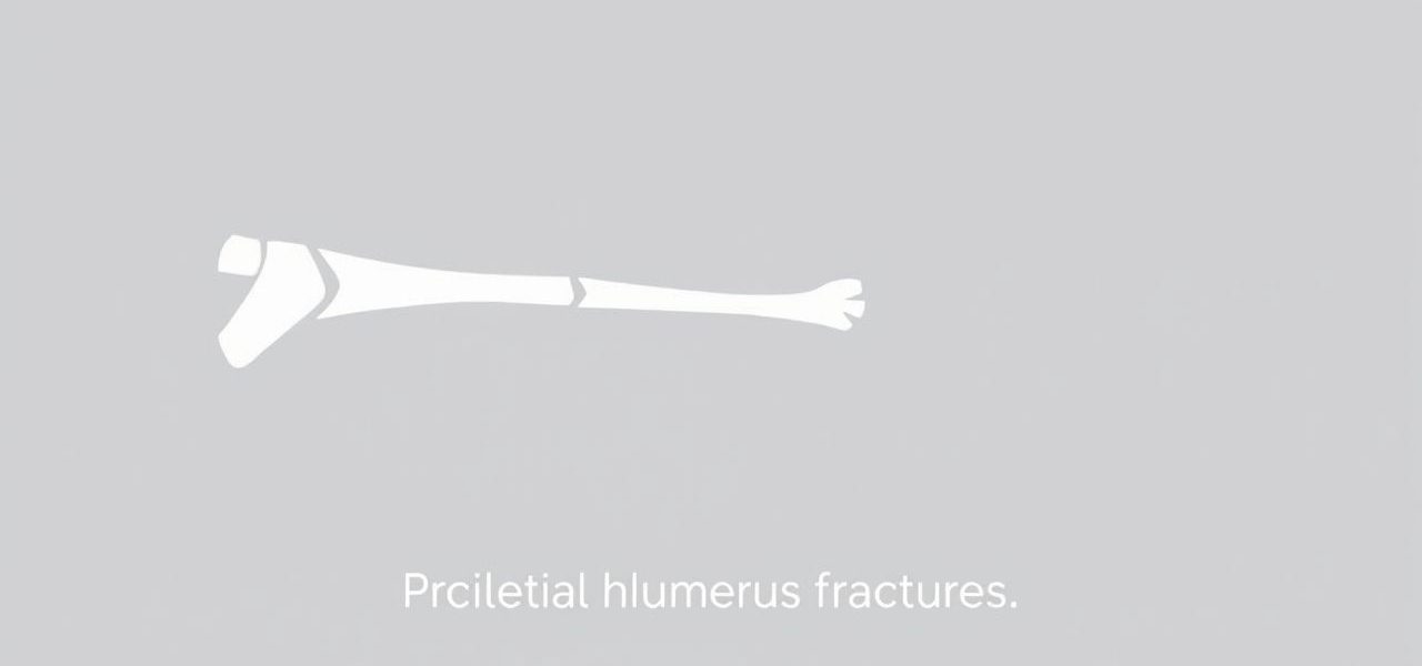
The distal humerus, located at the lower end of the upper arm bone, plays a crucial role in elbow movement and arm stability. Fractures in this region can significantly impact daily activities, especially those involving bending or straightening the elbow. Because of the complexity of the bone’s anatomy and its relationship to surrounding nerves and blood vessels, understanding the classification of distal humerus fractures is essential for diagnosis, treatment planning, and recovery. Proper classification helps surgeons determine whether a fracture requires surgical fixation, conservative management, or specialized rehabilitation. It also improves communication between healthcare professionals when discussing the severity and type of injury.
Anatomy of the Distal Humerus
The distal humerus forms the upper part of the elbow joint and consists of several important structures. These include the medial and lateral condyles, the trochlea, and the capitellum, which articulate with the forearm bones the ulna and radius. Between these bony landmarks are fossae that allow for elbow flexion and extension. The ulnar nerve runs close to the medial side, making it vulnerable to injury during fractures or surgical repair.
Due to its complex shape, fractures of the distal humerus often involve multiple fragments and can extend into the joint surface, complicating treatment.
Importance of Classification
Classification of distal humerus fractures allows orthopedic surgeons to assess fracture patterns, predict possible complications, and decide the most effective surgical or nonsurgical approach. A clear classification system also helps in research and outcome comparison across different patient groups and treatment techniques.
Main Classification Systems
Several classification systems are used to describe distal humerus fractures, each offering a different perspective on fracture pattern and severity. The most common systems include the AO/OTA classification and the Jupiter classification. Each method focuses on fracture location, involvement of the joint surface, and the presence of comminution (multiple bone fragments).
AO/OTA Classification
The AO/OTA (Arbeitsgemeinschaft für Osteosynthesefragen/Orthopaedic Trauma Association) system is one of the most widely used methods for categorizing distal humerus fractures. It divides fractures into three main types, each with subcategories.
- Type A – Extra-articular fractures
- A1 Simple extra-articular fracture without comminution
- A2 Extra-articular fracture with simple metaphyseal involvement
- A3 Comminuted extra-articular fracture
- Type B – Partial articular fractures
- B1 Lateral condyle fracture
- B2 Medial condyle fracture
- B3 Fractures involving the trochlea
- Type C – Complete articular fractures
- C1 Simple articular fracture with simple metaphyseal involvement
- C2 Simple articular fracture with metaphyseal comminution
- C3 Comminuted articular and metaphyseal fracture
This classification is particularly valuable because it specifies whether the fracture involves the joint surface and how complex the break is, which greatly influences treatment decisions.
Jupiter Classification
The Jupiter classification system focuses on fracture patterns and the involvement of the columns of the distal humerus. The distal humerus is often described as having two columns medial and lateral that provide structural support to the elbow joint. This system categorizes fractures as
- Unicolumn fractures– Involving only the medial or lateral column
- Bicolumn fractures– Involving both columns but may or may not include the articular surface
- Intercondylar fractures– Involving both columns and extending into the joint space
This classification emphasizes the structural stability of the elbow and helps guide surgical reconstruction approaches that restore both columns for optimal joint function.
Mechanisms of Injury
Understanding how distal humerus fractures occur also assists in classification and treatment planning. Common causes include
- Direct trauma, such as a fall onto a flexed elbow
- High-energy impacts, such as in motor vehicle accidents
- Low-energy falls in elderly patients with osteoporosis
The energy level of the injury often correlates with the degree of comminution and displacement seen on imaging studies.
Role of Imaging in Classification
Accurate classification depends on high-quality imaging. Standard X-rays in anteroposterior and lateral views are essential, but computed tomography (CT) scans provide a more detailed view of complex fractures, especially those extending into the joint. CT with 3D reconstruction allows surgeons to visualize fracture lines and fragment positions, making classification more precise.
Challenges in Classification
While classification systems provide structure, some fractures are difficult to categorize due to unusual patterns or overlapping characteristics. Factors that complicate classification include
- Severe comminution obscuring anatomical landmarks
- Combined injuries, such as elbow dislocation with fracture
- Pre-existing bone deformities or prior surgeries
In such cases, a combination of classification systems and descriptive terms may be used to fully explain the injury.
Impact on Treatment Decisions
The classification of distal humerus fractures directly influences treatment planning. For example
- Type A fractures (AO/OTA) may be treated conservatively if stable and well-aligned
- Type B fractures often require fixation to restore joint congruity
- Type C fractures almost always require surgical intervention with plate fixation or other reconstructive techniques
Similarly, Jupiter’s bicolumn and intercondylar fractures typically need dual plate fixation to ensure adequate stability for early motion and to minimize stiffness.
Rehabilitation Considerations
Regardless of fracture type, early controlled movement is important to prevent elbow stiffness, one of the most common complications after distal humerus fractures. Rehabilitation protocols depend on fracture stability and the type of surgical fixation used. Stable extra-articular fractures may allow earlier range-of-motion exercises, while complex articular fractures may require longer periods of immobilization before rehabilitation begins.
Prognosis and Long-Term Outcomes
Classification also helps predict functional outcomes. Simple extra-articular fractures tend to heal with minimal loss of motion, while complex comminuted articular fractures carry a higher risk of residual stiffness, post-traumatic arthritis, and decreased strength. Surgical expertise, patient age, and adherence to rehabilitation protocols all influence recovery.
The classification of distal humerus fractures is a critical step in managing these complex injuries. Systems like AO/OTA and Jupiter provide structured approaches to describing fracture patterns, guiding treatment, and predicting outcomes. By understanding the anatomy, mechanisms of injury, and imaging findings, healthcare providers can classify fractures accurately and choose the most effective management strategy, ultimately improving patient recovery and long-term elbow function.