An X-ray of the nasopharynx in a lateral view is a specialized imaging technique used to evaluate the structures of the upper airway, including the nasal cavity, pharynx, and surrounding tissues. This type of X-ray provides a side-on perspective, which helps radiologists and clinicians assess anatomical details that may not be visible in standard frontal views. It is commonly used to investigate conditions such as infections, tumors, congenital abnormalities, and obstructions that affect breathing, swallowing, or speech. The lateral view allows for precise measurement of structures and provides a valuable tool for diagnosis and treatment planning in otolaryngology and general medicine.
Understanding the Nasopharynx
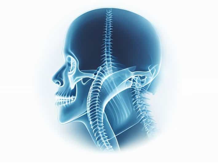
The nasopharynx is the uppermost section of the pharynx, located behind the nasal cavity and above the soft palate. It serves as a passageway for air during breathing and connects the nasal passages to the oropharynx. The nasopharynx also houses important structures, such as the pharyngeal tonsils (adenoids) and openings of the Eustachian tubes, which help regulate pressure in the middle ear. Because of its complex anatomy and proximity to vital structures, precise imaging is crucial for diagnosing abnormalities in this region. The lateral view X-ray provides clear visualization of the nasopharynx, soft tissues, and surrounding bony landmarks.
Purpose of Lateral View X-Ray
The lateral view X-ray of the nasopharynx is performed to
- Assess the size and shape of the adenoids and pharyngeal space.
- Identify obstructions in the airway caused by enlarged tissues or masses.
- Detect structural abnormalities, including congenital deformities or trauma-related changes.
- Monitor chronic conditions such as adenoid hypertrophy or nasopharyngeal carcinoma.
- Assist in preoperative planning for procedures like adenoidectomy or other ENT surgeries.
Procedure and Technique
Performing a nasopharynx lateral view X-ray involves specific positioning and technical considerations to ensure accurate imaging. The patient is usually asked to stand or sit upright with their head in a neutral position. The X-ray beam is directed from one side of the head to the other, capturing a lateral projection that highlights the nasopharynx, posterior pharyngeal wall, cervical spine, and surrounding soft tissues. Radiologic technologists may use markers or reference points to measure airway space, tissue thickness, and anatomical relationships. Proper positioning is essential for minimizing distortion and obtaining a diagnostic-quality image.
Patient Preparation
Preparation for a lateral nasopharynx X-ray is generally minimal but important for optimal results. Patients may be asked to remove jewelry, glasses, or hair accessories that could interfere with imaging. Instructions often include keeping the head still and maintaining normal breathing during the exposure. Children may require additional support or immobilization to ensure accurate positioning. The procedure is quick, typically taking only a few minutes, and is non-invasive, causing minimal discomfort.
Interpretation of Results
Radiologists examine the lateral view X-ray to assess both soft tissue and bony structures. Key aspects evaluated include the adenoid size, airway patency, alignment of cervical vertebrae, and any abnormal masses or lesions. Measurements such as the adenoid-nasopharyngeal ratio can help determine the degree of airway obstruction, which is particularly useful in pediatric patients with recurrent respiratory issues or sleep disturbances. Abnormal findings may indicate the need for further evaluation using additional imaging modalities, such as CT or MRI, for more detailed assessment.
Common Indications
Some common indications for performing a lateral view X-ray of the nasopharynx include
- Chronic nasal obstruction or difficulty breathing through the nose.
- Recurrent ear infections or suspected Eustachian tube dysfunction.
- Snoring, sleep apnea, or other sleep-related breathing disorders.
- Suspected tumors or masses in the nasopharyngeal region.
- Assessment of adenoid hypertrophy in children causing speech or swallowing difficulties.
Advantages and Limitations
The lateral view X-ray of the nasopharynx offers several advantages. It is quick, widely available, cost-effective, and provides a clear image of airway structures and soft tissues. This imaging method is particularly useful in pediatric populations for evaluating adenoids and detecting obstructions without the need for more invasive procedures. However, limitations exist. Overlapping structures, patient movement, and variations in positioning can affect image clarity. Additionally, X-rays provide limited soft tissue contrast compared to CT or MRI, so further imaging may be necessary for complex cases or suspected tumors.
Safety Considerations
Like all X-rays, lateral nasopharynx imaging involves exposure to a small amount of ionizing radiation. The dose is generally low, and radiologists take precautions to minimize exposure, such as using lead shields and limiting the field of view. Pregnant patients or individuals requiring multiple imaging studies should discuss potential risks with their healthcare provider. Modern digital X-ray systems have further reduced radiation exposure while improving image quality, making the procedure safe for most patients, including children.
Clinical Significance
The lateral view X-ray of the nasopharynx is a valuable diagnostic tool for ENT specialists, pediatricians, and general practitioners. It allows for the early detection of abnormalities, guides treatment decisions, and assists in monitoring response to therapy. Accurate imaging contributes to improved patient outcomes, particularly in conditions like adenoid hypertrophy, chronic sinusitis, and nasopharyngeal masses. It also plays a role in surgical planning by providing detailed anatomical information critical for procedures in the upper airway.
An X-ray of the nasopharynx in a lateral view is a simple yet powerful imaging technique that provides essential information about the upper airway and surrounding structures. By offering a clear side-on perspective, it enables radiologists to detect abnormalities, assess airway patency, and support diagnosis and treatment planning. The procedure is safe, quick, and widely accessible, making it an indispensable tool in otolaryngology and general medical practice. For patients experiencing nasal obstruction, recurrent infections, or suspected masses, the lateral nasopharynx X-ray serves as a critical first step in evaluation and management, combining efficiency with diagnostic accuracy.