The kidney and ureter are essential organs in the human urinary system that work together to filter blood, remove waste, and transport urine to the bladder. Understanding the anatomy of the kidney and ureter provides important insight into how the body maintains fluid balance and eliminates toxins. These organs play a vital role not only in excretion but also in regulating electrolytes, blood pressure, and red blood cell production. Their precise structure and coordination are crucial for maintaining homeostasis and supporting overall health.
Gross Anatomy of the Kidney
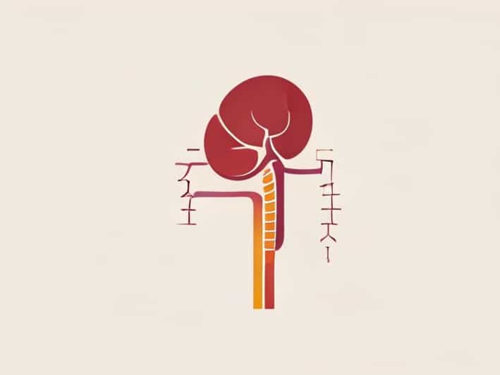
The kidneys are two bean-shaped organs located in the posterior abdominal cavity on either side of the spine, usually between the levels of the twelfth thoracic (T12) and third lumbar (L3) vertebrae. The right kidney sits slightly lower than the left due to the position of the liver. Each kidney measures about 10 12 centimeters in length, 5 7 centimeters in width, and weighs approximately 150 grams in adults.
External Structure
The kidney is enclosed by three layers of protective tissue. The innermost layer, the renal capsule, is a tough fibrous covering that directly surrounds the kidney. Outside the capsule lies a layer of adipose tissue called the perirenal fat, which cushions and protects the organ from trauma. The outermost layer, the renal fascia, anchors the kidney to surrounding structures and maintains its position in the abdomen.
The lateral surface of each kidney is convex, while the medial surface is concave and features a central depression known as the hilum. The hilum serves as the entry and exit point for the renal artery, renal vein, lymphatics, and ureter. This region connects to the renal sinus, a cavity within the kidney that houses blood vessels and the renal pelvis.
Internal Structure
The kidney’s internal anatomy is divided into two main regions the renal cortex and the renal medulla. The renal cortex forms the outer portion and contains the renal corpuscles and convoluted tubules, which are essential for filtration and absorption. The medulla lies beneath the cortex and is organized into 8 18 triangular structures called renal pyramids.
Each pyramid terminates in a renal papilla that projects into a minor calyx. These minor calyces merge to form major calyces, which in turn combine to create the renal pelvis a funnel-shaped structure that collects urine and channels it into the ureter. This precise organization facilitates the flow of filtered urine from the microscopic nephrons to the collecting ducts and eventually to the bladder.
Microscopic Anatomy of the Kidney
The functional unit of the kidney is the nephron, and each kidney contains approximately one million nephrons. These microscopic structures perform the essential tasks of filtration, reabsorption, secretion, and excretion.
Structure of a Nephron
Each nephron consists of two primary components the renal corpuscle and the renal tubule. The renal corpuscle includes the glomerulus a network of capillaries surrounded by Bowman’s capsule. Blood enters the glomerulus through the afferent arteriole, and filtration occurs as plasma passes through the glomerular membrane into Bowman’s space.
The filtrate then travels through the renal tubule, which is composed of the following segments
- Proximal convoluted tubule (PCT)Responsible for reabsorbing water, glucose, amino acids, and ions back into the bloodstream.
- Loop of HenleExtends into the medulla and establishes a concentration gradient that helps concentrate urine.
- Distal convoluted tubule (DCT)Regulates ion exchange and responds to hormones such as aldosterone for sodium and potassium balance.
- Collecting ductReceives urine from multiple nephrons and fine-tunes water reabsorption under the influence of antidiuretic hormone (ADH).
The nephrons collectively ensure that waste products such as urea, creatinine, and uric acid are excreted while conserving essential substances like glucose and electrolytes.
Blood Supply of the Kidney
The kidneys receive approximately 20 25% of cardiac output, highlighting their critical role in blood filtration. Blood enters the kidney through the renal artery, which branches off from the abdominal aorta. The renal artery divides into segmental, interlobar, arcuate, and interlobular arteries, ultimately leading to afferent arterioles that supply the glomeruli.
After filtration, blood exits the glomerulus through the efferent arteriole. These vessels form a network of peritubular capillaries around the renal tubules, allowing for reabsorption and secretion. Blood then returns to the general circulation via the interlobular veins, arcuate veins, and renal vein, which drains into the inferior vena cava.
Overview of the Ureter
The ureter is a muscular tube approximately 25 30 centimeters long that transports urine from the renal pelvis to the urinary bladder. There are two ureters one for each kidney. Each ureter follows a downward path along the posterior abdominal wall, passing over the pelvic brim before entering the bladder at its posterolateral surface.
Layers of the Ureter
The ureter wall is composed of three layers that allow it to carry urine efficiently through peristaltic movements
- MucosaThe innermost layer, lined with transitional epithelium, allows the ureter to stretch as urine passes through.
- MuscularisThis middle layer consists of inner longitudinal and outer circular smooth muscle layers. Their rhythmic contractions propel urine toward the bladder.
- AdventitiaThe outermost connective tissue layer anchors the ureter to surrounding structures and provides vascular support.
These layers work together to ensure a one-way flow of urine, preventing backflow and maintaining kidney health.
Constriction Points
There are three natural narrowings along the ureter where kidney stones are most likely to become lodged
- At the junction between the renal pelvis and the ureter (ureteropelvic junction).
- As the ureter crosses over the pelvic brim near the iliac vessels.
- At the point where the ureter enters the bladder wall (ureterovesical junction).
Understanding these constriction points is important in diagnosing and treating conditions such as ureteral obstruction and renal colic caused by kidney stones.
Physiological Connection Between Kidney and Ureter
The kidney and ureter work together to ensure that urine formed in the renal nephrons is efficiently transported to the bladder for storage. Once the collecting ducts in the kidney deliver urine to the renal pelvis, peristaltic contractions begin in the ureter to move it downward. This movement is autonomous and continues even if the ureter is detached from the kidney, demonstrating the organ’s independent contractile function.
The junction between the kidney and the ureter, called the ureteropelvic junction, acts as a valve-like region that prevents backflow. Proper coordination between renal filtration and ureteral transport is vital to prevent hydronephrosis, a condition where urine backs up into the kidney, causing swelling and potential damage.
Common Disorders Affecting the Kidney and Ureter
Several medical conditions can affect the anatomy and function of the kidney and ureter. Some common disorders include
- Kidney stones (nephrolithiasis)Hard deposits formed from minerals and salts that can obstruct the ureter.
- HydronephrosisSwelling of the kidney due to urine buildup, often caused by ureteral blockage.
- PyelonephritisA bacterial infection of the kidney that can spread from the ureter or bladder.
- Ureteral stricturesNarrowing of the ureter that interferes with urine flow.
- Congenital anomaliesConditions like duplicated ureters or ectopic ureteral openings that alter normal urinary drainage.
Early diagnosis and imaging techniques such as ultrasound, CT scans, and MRI are essential for identifying these conditions and ensuring proper treatment.
The anatomy of the kidney and ureter reveals a remarkable system of structure and function that ensures the body efficiently removes waste while maintaining internal balance. The kidneys’ intricate nephron network filters blood with precision, and the ureters’ muscular structure delivers urine reliably to the bladder. Any disruption in their anatomy or physiology can lead to significant health issues, emphasizing the importance of understanding and caring for these vital organs. Through a combination of protective layers, complex vasculature, and synchronized activity, the kidney and ureter exemplify the human body’s extraordinary ability to regulate and sustain life.