The anatomy of the ureter in females is a fascinating aspect of the urinary system that highlights how the body efficiently manages waste removal while maintaining harmony with nearby reproductive organs. Understanding the structure, pathway, and relations of the female ureter is essential for students of anatomy, healthcare professionals, and anyone interested in how the human body functions. The female ureter’s location is of particular importance in gynecology and urology, as its close association with the uterus, vagina, and other pelvic organs makes it susceptible to injury during certain medical procedures.
Overview of the Ureter
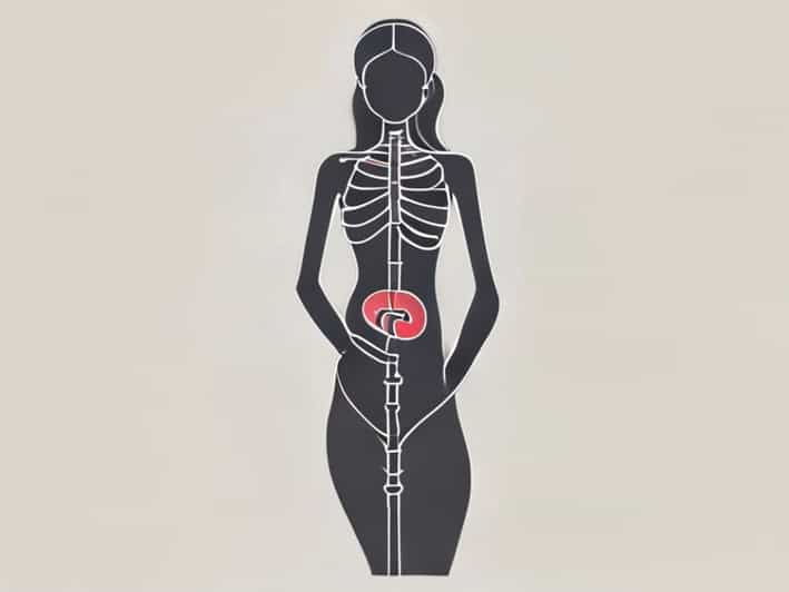
The ureter is a paired muscular tube that carries urine from the kidneys to the urinary bladder. Each kidney has one ureter, making two in total. The ureter functions as a conduit that propels urine through peristaltic contractions wave-like muscle movements that ensure urine flows smoothly toward the bladder. In females, the ureter measures approximately 25 to 30 centimeters in length and follows a slightly different course compared to males because of the presence of reproductive organs such as the uterus and ovaries.
Gross Anatomy of the Female Ureter
The female ureter can be divided into two major parts the abdominal part and the pelvic part. These sections differ in terms of anatomical relationships and clinical significance.
1. Abdominal Part of the Ureter
The abdominal part of the ureter extends from the renal pelvis (the funnel-shaped part of the kidney) to the pelvic brim. It descends along the anterior surface of the psoas major muscle, just behind the peritoneum. The ureter crosses important structures, such as the gonadal vessels, as it moves downward. In the female body, it lies slightly lateral to the ovarian artery and vein. This portion of the ureter receives its blood supply from the renal arteries, gonadal arteries, and aortic branches.
2. Pelvic Part of the Ureter
Once the ureter reaches the pelvic brim, it enters the pelvic cavity by crossing the bifurcation of the common iliac artery. The pelvic part is clinically significant because it passes near the reproductive organs. In females, it runs along the lateral pelvic wall, under the uterine artery, and then curves forward and medially toward the bladder. This relationship is famously remembered by the phrase water under the bridge, meaning the ureter (water) passes under the uterine artery (the bridge).
Course of the Ureter in the Female Pelvis
The female ureter’s journey through the pelvis is complex due to its proximity to reproductive organs. It travels through several anatomical regions before reaching the bladder.
- At the pelvic brimThe ureter crosses the common iliac artery and vein, entering the lesser pelvis.
- Along the lateral pelvic wallIt runs within the connective tissue of the broad ligament, close to the ovary and uterine vessels.
- Near the cervixThe ureter passes approximately 1.5 2 centimeters lateral to the cervix, where it is in close relation to the uterine artery.
- Before entering the bladderThe ureter pierces the bladder wall obliquely, forming the ureteric orifice on the posterior aspect of the bladder (trigone area).
Relations of the Female Ureter
The anatomical relations of the female ureter are crucial, especially during surgical procedures such as hysterectomy or cesarean section. Any accidental injury to the ureter can lead to serious complications like urinary leakage or obstruction. Understanding these relationships helps surgeons avoid such risks.
Anterior Relations
In the abdominal region, the ureter lies behind the peritoneum and is crossed by the ovarian vessels. In the pelvis, the anterior relations include the ovary, uterine artery, and the base of the broad ligament. Near the bladder, it lies anterior to the vagina’s upper part.
Posterior Relations
Posteriorly, the ureter rests on the psoas major muscle in the abdomen and on the internal iliac vessels within the pelvis. This relationship provides stability and helps maintain the ureter’s orientation as it travels downward.
Lateral Relations
Laterally, the ureter is related to the parietal peritoneum and pelvic fascia. In the lower pelvis, it is closely associated with the uterosacral ligament and the pelvic wall.
Medial Relations
Medially, the ureter comes close to the uterus and the cervix, especially where it passes beneath the uterine artery. This intimate relationship makes it particularly vulnerable during gynecologic surgeries involving the uterine or cervical region.
Histological Structure of the Ureter
The wall of the ureter is composed of three main layers, each playing a vital role in the transport of urine
- MucosaThe inner lining is made of transitional epithelium (urothelium), which allows the ureter to stretch as urine passes through. The mucosa also contains folds that help prevent backflow.
- Muscular layerThis middle layer consists of smooth muscle arranged in two main layers an inner longitudinal layer and an outer circular layer. In the lower third of the ureter, an additional outer longitudinal layer appears, strengthening peristaltic contractions.
- AdventitiaThe outermost layer contains connective tissue, blood vessels, lymphatics, and nerves. It anchors the ureter to surrounding structures and provides vascular support.
Blood Supply and Lymphatic Drainage
The female ureter receives a segmental blood supply from several nearby arteries. The upper portion is supplied by the renal arteries, the middle part by the gonadal arteries, and the lower part by branches of the uterine and vaginal arteries. Venous drainage follows a similar pattern, emptying into corresponding veins.
Lymph from the ureter drains into regional lymph nodes, including the lumbar and internal iliac nodes. This drainage pattern is important in the spread of infections or malignancies affecting the urinary or reproductive systems.
Nerve Supply of the Ureter
The ureter’s nerve supply comes from the renal, aortic, hypogastric, and pelvic plexuses. These nerves contain both sympathetic and parasympathetic fibers. Sympathetic nerves help regulate peristalsis and constrict the ureter, while parasympathetic fibers assist in relaxation and coordination of urine flow. Pain from the ureter is often referred to the lower abdomen, flank, or inner thigh, depending on the affected segment.
Functional Role of the Female Ureter
The ureter’s main role is to transport urine from the kidneys to the bladder without allowing backflow. The oblique entry of the ureter into the bladder wall acts as a natural valve that prevents urine from re-entering the ureter during bladder contraction. This mechanism is essential for preventing urinary tract infections and maintaining normal kidney function.
Clinical Significance
Due to its proximity to the uterus and cervix, the female ureter is at risk during surgeries like hysterectomy or pelvic organ repair. Accidental ligation or transection can lead to complications such as ureteral obstruction, fistula formation, or urinary leakage. Radiological imaging, such as ultrasound or CT urograms, helps detect obstructions or stones within the ureter.
Ureteral stones, infections, and congenital abnormalities can also affect the ureter’s function. Understanding its anatomy allows physicians to plan safe surgical approaches and provide accurate diagnoses.
The anatomy of the ureter in females reveals an intricate balance between the urinary and reproductive systems. Its close relationship with organs like the uterus, ovaries, and vagina makes it both functionally efficient and clinically significant. From its origin at the renal pelvis to its termination in the bladder, the ureter demonstrates a remarkable adaptation to ensure proper urine flow while coexisting with vital reproductive structures. A deep understanding of its pathway, relations, and structure is essential for healthcare professionals involved in pelvic surgeries, urology, and gynecology. Ultimately, the female ureter’s anatomy reflects the complexity and precision of human physiology, where even small structures play vital roles in maintaining overall health.