The human foot is a marvel of engineering, supporting the weight of the body while allowing for balance, movement, and shock absorption. One key part of this complex structure is the tarsus, a cluster of bones that forms the rear portion of the foot, also known as the hindfoot and midfoot. These bones play an essential role in locomotion, offering strength and flexibility for walking, running, and jumping. Understanding the bones of the tarsus gives insight into how the foot functions as a whole and how it connects to other parts of the skeletal system.
What Is the Tarsus?
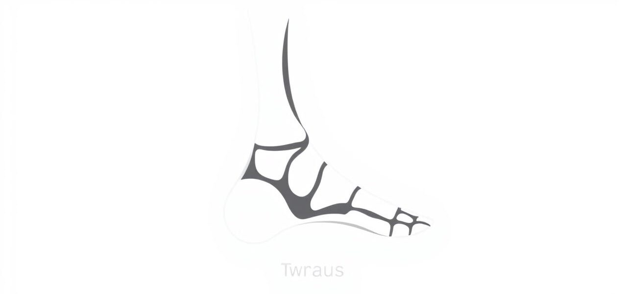
The tarsus refers to a group of seven irregularly shaped bones located in the posterior part of the foot, between the lower leg bones (tibia and fibula) and the metatarsals of the midfoot. These bones are responsible for forming the ankle and part of the arch of the foot. They are larger and stronger than the bones of the forefoot, reflecting their role in weight-bearing and stability.
Overview of the Seven Tarsal Bones
The seven bones of the tarsus are:
- Talus
- Calcaneus
- Navicular
- Cuboid
- Medial cuneiform
- Intermediate cuneiform
- Lateral cuneiform
Each of these tarsal bones serves a specific purpose and connects with surrounding bones to form joints that allow for a range of foot movements.
The Talus: The Keystone of the Ankle
The talus is the second largest of the tarsal bones and sits on top of the calcaneus. It forms the lower part of the ankle joint by articulating with the tibia and fibula. Unlike most bones in the body, the talus does not have any muscle attachments. Instead, it serves as a critical connector, transferring weight and movement from the leg to the foot.
Function of the Talus
The talus plays a central role in foot and ankle motion. It allows the foot to move up and down (dorsiflexion and plantarflexion) and contributes to side-to-side movements. Damage to the talus, such as fractures or arthritis, can significantly impair mobility.
The Calcaneus: The Heel Bone
The calcaneus is the largest tarsal bone and forms the heel. It lies beneath the talus and supports much of the body’s weight during standing and walking. The calcaneus also serves as the attachment site for the Achilles tendon, the strongest tendon in the human body.
Role in Movement
The calcaneus acts as a lever arm for the calf muscles. It absorbs impact during heel strikes and helps propel the body forward. It forms the subtalar joint with the talus, which is essential for foot inversion and eversion.
The Navicular Bone: A Midfoot Stabilizer
Situated in front of the talus, the navicular bone connects the rearfoot to the midfoot. It gets its name from its boat-like shape. The navicular articulates with the talus and the three cuneiform bones.
Function of the Navicular
This bone plays a key role in maintaining the arch of the foot. Ligaments and tendons, including the posterior tibial tendon, attach to the navicular, helping stabilize the medial side of the foot. Injury or dysfunction in this area can lead to flatfoot or arch problems.
The Cuboid Bone: Lateral Support
The cuboid bone is located on the lateral (outer) side of the foot. It articulates with the calcaneus, the fourth and fifth metatarsals, and the lateral cuneiform.
Structural Role
The cuboid supports the lateral column of the foot. It also has a groove that allows passage for the peroneus longus tendon, which assists in arch support and foot stabilization. Injuries to the cuboid, such as cuboid syndrome, can cause lateral foot pain and difficulty walking.
The Three Cuneiform Bones
The cuneiform bones medial, intermediate, and lateral are wedge-shaped and located in the midfoot between the navicular and the first three metatarsals. They help form the transverse arch of the foot and contribute to its shape and flexibility.
Functions of the Cuneiforms
- Medial cuneiform: The largest of the three, it supports the first metatarsal and plays a vital role in maintaining the medial arch.
- Intermediate cuneiform: Smaller and centrally located, it connects the second metatarsal to the navicular.
- Lateral cuneiform: Articulates with the third metatarsal and both the intermediate cuneiform and cuboid.
These bones serve as connection points for muscles and ligaments, and their alignment is crucial for proper foot mechanics.
Joints Formed by the Tarsal Bones
The bones of the tarsus form several joints that enable complex foot movements and flexibility:
- Ankle joint: Formed by the talus, tibia, and fibula.
- Subtalar joint: Between the talus and calcaneus, allowing rotation and tilting of the foot.
- Transverse tarsal joint: Includes the talonavicular and calcaneocuboid joints, important for adapting to different surfaces.
- Tarsometatarsal joints: Connect the tarsals to the metatarsals, enabling flexion and extension.
These joints allow the foot to absorb shock, adjust to uneven terrain, and assist in propulsion during movement.
Common Conditions Affecting the Tarsal Bones
The tarsal bones, while strong, can be affected by a range of conditions due to their role in bearing weight and enabling movement. Some of the most common problems include:
- Tarsal coalition: A congenital condition where two or more tarsal bones are fused, reducing mobility and causing pain.
- Stress fractures: Especially in the navicular and talus due to repetitive impact in athletes.
- Arthritis: Degeneration in the joints between tarsal bones, leading to stiffness and pain.
- Flatfoot: Collapse of the arch involving dysfunction of the navicular and associated tendons.
Treatment for tarsal bone issues ranges from rest and orthotics to surgery, depending on the severity and cause.
Importance of the Tarsus in Biomechanics
The bones of the tarsus form the foundation of the foot’s structure and function. They work together to support body weight, maintain balance, and adapt to various movements. The arch system, made possible by these bones, allows the foot to function like a spring, storing and releasing energy efficiently during locomotion.
Adaptability and Strength
The tarsal bones must be both flexible and strong. Their alignment and condition influence posture, gait, and the health of other joints, including the knees, hips, and spine. Understanding the role of each tarsal bone helps in diagnosing foot pain and creating effective treatment plans.
The bones of the tarsus talus, calcaneus, navicular, cuboid, and the three cuneiforms are essential components of the foot’s structure. Each bone has a unique role in mobility, support, and flexibility. Together, they form a dynamic system that enables the foot to function properly during a wide range of activities. Whether in everyday walking or high-impact sports, the tarsal bones play a crucial role in maintaining balance and performance. Knowing the anatomy and function of these bones not only helps in understanding the complexity of the human foot but also in recognizing the importance of foot health in overall wellbeing.