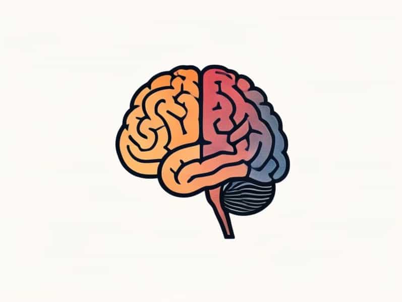
The human brain is an extraordinarily complex organ that serves as the control center for all bodily functions, cognition, and behavior. One of the key aspects of understanding how the brain works is studying its different cortices. The cerebral cortex, the outermost layer of the brain, is divided into multiple specialized regions that handle sensory input, motor control, cognition, and higher-order thinking. Learning about the different cortices of the brain allows researchers, students, and medical professionals to understand how various brain areas contribute to everyday functions such as movement, perception, language, memory, and decision-making.
Overview of the Cerebral Cortex
The cerebral cortex is a highly folded structure that increases the surface area of the brain, allowing for greater cognitive capacity. It is divided into two hemispheres the left and right connected by the corpus callosum, which enables communication between them. Each hemisphere is further subdivided into lobes, each containing specific cortical areas responsible for distinct functions. The different cortices of the brain include the frontal, parietal, temporal, and occipital cortices, as well as specialized areas such as the insular cortex and the cingulate cortex.
Frontal Cortex
The frontal cortex, located at the front of the brain, is involved in a wide range of functions associated with higher cognitive abilities. It plays a critical role in voluntary movement, decision-making, problem-solving, and social behavior. The frontal cortex can be divided into several important subregions
Prefrontal Cortex
- Responsible for executive functions such as planning, reasoning, and goal setting.
- Involved in regulating social behavior and personality expression.
- Crucial for working memory and attention control.
Primary Motor Cortex
- Located in the precentral gyrus of the frontal lobe.
- Controls voluntary movements of specific body parts.
- Organized somatotopically, meaning different regions correspond to different body areas.
Parietal Cortex
The parietal cortex, situated behind the frontal cortex, processes sensory information and integrates it with motor commands. This area is essential for spatial awareness, perception of touch, and coordination of movements in response to sensory input.
Primary Somatosensory Cortex
- Located in the postcentral gyrus of the parietal lobe.
- Receives tactile information such as pressure, texture, and temperature.
- Also involved in proprioception, which is the perception of body position in space.
Posterior Parietal Cortex
- Integrates sensory input with motor commands to guide actions.
- Plays a role in spatial attention, navigation, and visual-motor coordination.
- Damage to this area can result in neglect syndrome or difficulty perceiving spatial relationships.
Temporal Cortex
The temporal cortex is located on the sides of the brain near the temples and is primarily responsible for auditory processing, language comprehension, and memory formation. It contains several important subregions
Primary Auditory Cortex
- Located in the superior temporal gyrus.
- Processes sound information such as pitch, volume, and rhythm.
- Essential for recognizing and interpreting auditory stimuli.
Wernicke’s Area
- Typically found in the left hemisphere’s superior temporal gyrus.
- Responsible for language comprehension and understanding spoken words.
- Damage can lead to Wernicke’s aphasia, characterized by impaired comprehension.
Medial Temporal Lobe
- Includes the hippocampus, critical for memory formation and retrieval.
- Supports learning and the consolidation of short-term memories into long-term memories.
Occipital Cortex
The occipital cortex, located at the back of the brain, is primarily responsible for visual processing. It interprets information received from the eyes and translates it into images and spatial perception. The key subregions include
Primary Visual Cortex
- Located in the calcarine sulcus.
- Receives input from the retina via the thalamus.
- Responsible for basic visual processing such as detecting shapes, colors, and movement.
Visual Association Areas
- Surround the primary visual cortex.
- Responsible for interpreting complex visual stimuli, recognizing objects, faces, and motion patterns.
- Damage to these areas can cause visual agnosia, an inability to recognize objects despite intact vision.
Insular Cortex
The insular cortex lies deep within the lateral sulcus and is involved in diverse functions, including emotion, taste perception, and interoception the perception of internal bodily states. It integrates sensory information with emotional and motivational states, contributing to feelings such as disgust, empathy, and self-awareness.
Cingulate Cortex
The cingulate cortex forms a part of the limbic system, running along the medial aspect of the brain. It plays a crucial role in emotion regulation, decision-making, attention, and pain perception. Subregions include the anterior cingulate cortex, which is involved in cognitive control and error detection, and the posterior cingulate cortex, which contributes to memory retrieval and spatial orientation.
Functional Integration of Cortices
While each cortex has specialized functions, the brain operates as an integrated network. The cortices communicate through white matter tracts, enabling complex behaviors, sensory-motor coordination, and higher-order cognition. For example, reading a book requires the occipital cortex for visual processing, the temporal cortex for language comprehension, and the frontal cortex for attention and planning. This interconnectedness demonstrates the importance of understanding both individual cortices and their collaborative functions.
Clinical Significance
Knowledge of the different cortices of the brain is essential in neurology, psychology, and rehabilitation medicine. Lesions or damage to specific cortices can result in functional deficits such as
- Frontal cortex damage leading to impaired decision-making or motor deficits.
- Parietal cortex injury causing sensory loss or spatial neglect.
- Temporal cortex lesions affecting memory and language comprehension.
- Occipital cortex damage resulting in visual impairments or cortical blindness.
- Insular or cingulate cortex dysfunction impacting emotion regulation and perception of pain.
The different cortices of the brain each contribute to the extraordinary range of human abilities, from basic sensory perception to complex reasoning and emotion regulation. The frontal, parietal, temporal, and occipital cortices, along with specialized regions like the insular and cingulate cortices, work in concert to support behavior, cognition, and sensory-motor integration. Understanding these cortices is crucial not only for neuroscience and medicine but also for appreciating the remarkable complexity and adaptability of the human brain. By studying the functions and interconnections of these cortical regions, researchers and clinicians can better diagnose, treat, and rehabilitate patients with neurological conditions, while educators can improve learning strategies that align with brain function.
Ultimately, the cerebral cortex is a testament to the intricate architecture and functional specialization of the brain. Each cortex, whether responsible for movement, sensation, vision, hearing, or emotion, contributes to the seamless operation of the human mind. Awareness of the different cortices of the brain enhances both scientific knowledge and practical application, from healthcare to education and cognitive enhancement.