The human cranium is a complex and carefully structured part of the skull that plays a crucial role in protecting the brain and supporting the structures of the head. In basic anatomy lessons, the cranium is often divided into regions to make it easier to study and understand. One of the most important of these regions is the rearmost part of the cranium. Although it may not always receive as much attention as the frontal or facial areas, this posterior section is essential for protection, balance, and neurological function. Understanding the division of the cranium, especially the rearmost part, helps learners appreciate how structure and function work together in the human body.
Overview of the Cranium
The cranium is the upper part of the skull that encloses and protects the brain. It is made up of several bones that are joined together by immovable joints called sutures. These bones form a rigid but slightly flexible structure that shields the brain from injury.
Anatomically, the cranium is often divided into different regions based on position and function. These divisions help students and medical professionals describe locations accurately and understand how different parts of the skull contribute to overall head anatomy.
Main Divisions of the Cranium
The cranium can be broadly divided into several regions, including the frontal, parietal, temporal, and occipital areas. Each region corresponds to specific bones and serves particular purposes.
- Frontal region at the front of the skull
- Parietal regions on the upper sides
- Temporal regions near the ears
- Occipital region at the back
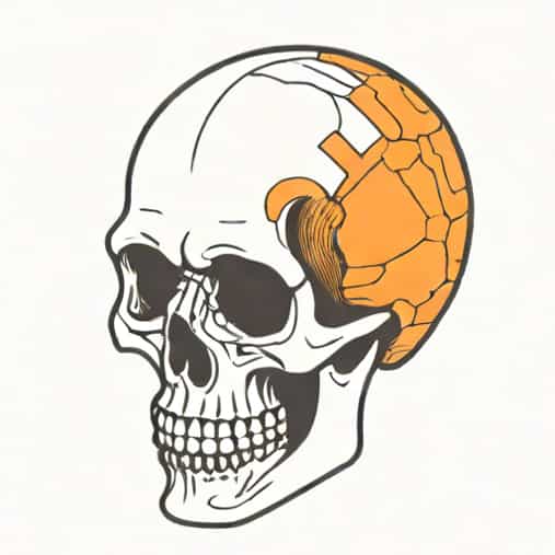
The rearmost part of the cranium is known as the occipital region, and it plays a key role in protecting vital brain structures.
The Occipital Region as the Rearmost Part
The rearmost part of the cranium is formed primarily by the occipital bone. This bone is located at the back and lower part of the skull and contributes to both the cranial vault and the base of the skull.
The occipital region protects the occipital lobes of the brain, which are responsible for processing visual information. Because of this function, damage to the rearmost part of the cranium can have serious effects on vision and coordination.
Location and Boundaries
The occipital bone is positioned behind the parietal bones and above the cervical vertebrae of the spine. It connects with surrounding bones through sutures, creating a strong and stable structure.
This region forms the back of the head and is easily felt as the rounded area at the lower rear of the skull.
Structure of the Occipital Bone
The occipital bone has a unique shape and several important features. One of its most significant structures is the foramen magnum, a large opening at the base of the skull.
The foramen magnum allows the spinal cord to pass through and connect with the brain. This opening makes the occipital bone essential not only for protection but also for communication between the brain and the rest of the body.
Key Anatomical Features
- Foramen magnum for spinal cord passage
- Occipital condyles for connection with the spine
- External occipital protuberance for muscle attachment
These features highlight how the rearmost part of the cranium supports both neurological and musculoskeletal functions.
Functions of the Rearmost Part of the Cranium
The primary function of the occipital region is protection. It shields the posterior part of the brain from external forces and trauma. Because the back of the head is vulnerable during falls or accidents, this protection is critical.
In addition to protection, the occipital bone supports head movement and posture. Its articulation with the first cervical vertebra allows the head to nod and rotate smoothly.
Role in Balance and Posture
The rearmost part of the cranium plays a role in maintaining balance and posture. Muscles attached to the occipital bone help stabilize the head and neck.
These muscles are especially active when maintaining an upright posture or performing movements such as looking up or turning the head.
Development of the Occipital Region
During fetal development, the occipital bone forms from several parts that gradually fuse together. In infants, these parts are separated by soft areas, allowing for brain growth.
As a person grows, the bones fuse completely, creating a solid and protective structure. This gradual development ensures both flexibility and safety during early life.
Clinical Importance of the Rearmost Cranium
The occipital region has important clinical significance. Injuries to the back of the head can affect vision, coordination, and consciousness. Medical professionals carefully assess this area in cases of head trauma.
Conditions such as fractures, tumors, or congenital abnormalities involving the occipital bone may impact the foramen magnum and spinal cord, making accurate diagnosis essential.
Common Medical Considerations
- Occipital bone fractures
- Compression near the foramen magnum
- Postural strain affecting occipital muscles
Understanding the anatomy of the rearmost part of the cranium helps in both prevention and treatment of these conditions.
Educational Importance in Anatomy
In basic anatomy education, learning the division of the cranium helps students organize complex information. Identifying the rearmost part as the occipital region makes it easier to relate structure to function.
This knowledge forms a foundation for more advanced studies in biology, medicine, and health sciences.
Relationship to Other Cranial Regions
The occipital region does not function in isolation. It works together with the parietal and temporal regions to form a complete protective shell for the brain.
The sutures connecting these bones allow the skull to absorb impact while maintaining overall strength. This cooperation between cranial regions highlights the efficiency of human skeletal design.
The division of the cranium helps simplify the study of skull anatomy, and the rearmost part plays a vital role within this structure. Known as the occipital region, it protects the back of the brain, supports head movement, and connects the brain to the spinal cord.
By understanding the anatomy and function of the rearmost part of the cranium, learners gain insight into how the human body protects its most important organ. This region may be at the back of the head, but its importance is central to vision, balance, and overall neurological health.