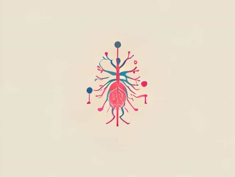
The development of the fetal autonomic nervous system (ANS) is one of the most fascinating and essential processes during pregnancy. It plays a crucial role in regulating involuntary functions such as heart rate, blood pressure, digestion, and respiratory control. Understanding how the fetal autonomic nervous system develops helps explain how a fetus adapts to its environment before birth and prepares for life outside the womb. This complex system begins forming early in gestation and continues to mature throughout pregnancy and even after delivery.
Overview of the Fetal Autonomic Nervous System
The autonomic nervous system is a division of the peripheral nervous system responsible for automatic bodily functions. It consists of two major components the sympathetic and parasympathetic nervous systems which work in balance to maintain homeostasis. In the fetus, the ANS begins to form as early as the fifth week of gestation, derived from neural crest cells that migrate to various parts of the developing body.
At this early stage, the foundations for reflexes and vital organ control are laid. The development of the fetal ANS is closely linked with the formation of the brainstem, spinal cord, and peripheral nerves. These neural pathways establish communication between the brain and vital organs, ensuring the fetus can respond to internal and external stimuli.
Stages of Fetal Autonomic Nervous System Development
First Trimester Formation of Neural Structures
During the first trimester, rapid cell division and differentiation occur. The neural tube forms by the fourth week and eventually develops into the brain and spinal cord. Neural crest cells migrate outward to form the sympathetic ganglia and parasympathetic pathways. These structures will later become responsible for regulating vital organ activity.
Although the basic framework of the autonomic nervous system is established during this period, it is not yet functional. The sympathetic nervous system develops slightly earlier than the parasympathetic branch, laying the groundwork for the body’s fight or flight mechanisms even before birth. However, true physiological responses do not appear until later in pregnancy.
Second Trimester Differentiation and Early Function
By the second trimester, around 13 to 27 weeks of gestation, the fetal autonomic nervous system begins to show signs of activity. Nerve fibers grow longer, synapses form, and neurotransmitters like norepinephrine and acetylcholine begin to appear. The sympathetic and parasympathetic systems start to differentiate more clearly.
At this stage, fetal heart rate patterns become an important indicator of autonomic activity. Variability in heart rate reflects the early balance between sympathetic and parasympathetic inputs. For example, a higher baseline heart rate suggests sympathetic dominance, while periods of slowing reflect parasympathetic modulation. These changes are often used in fetal monitoring to assess neurological and cardiovascular development.
Third Trimester Maturation and Integration
In the third trimester, from 28 weeks to birth, the fetal autonomic nervous system undergoes significant maturation. Neural connections between the brainstem and peripheral organs become more refined, allowing the fetus to regulate essential functions like breathing movements, circulation, and digestion.
Fetal heart rate variability increases, indicating that the parasympathetic system is becoming more active and balanced with the sympathetic branch. This balance is essential for proper physiological adaptation at birth. For instance, the ability to adjust heart rate and blood pressure helps the newborn transition from intrauterine to extrauterine life, where oxygen must be obtained through breathing instead of the placenta.
Sympathetic Nervous System Development
The sympathetic nervous system is responsible for preparing the body for action. It controls responses such as increased heart rate, dilation of airways, and redistribution of blood to muscles. In the fetus, sympathetic development begins around the sixth week of gestation and continues throughout pregnancy.
Sympathetic ganglia form along the spinal cord and connect to various organs. The adrenal medulla, an important part of the sympathetic system, begins producing catecholamines like adrenaline and noradrenaline during mid-gestation. These hormones help the fetus respond to stress, such as temporary hypoxia, and play a role in maintaining circulation.
- By 10 12 weeks Sympathetic fibers start connecting to the heart and blood vessels.
- By 20 weeks Adrenal medulla becomes functionally active, secreting catecholamines.
- By 30 weeks The fetus shows measurable sympathetic responses to stress or movement.
The sympathetic system’s early development is crucial because it ensures the fetus can adapt to physiological challenges. It also supports growth by regulating blood flow and nutrient distribution through vascular tone control.
Parasympathetic Nervous System Development
The parasympathetic nervous system promotes relaxation and energy conservation. It slows the heart rate, stimulates digestion, and aids in restorative processes. Its development follows the sympathetic system but continues into the late stages of pregnancy and even after birth.
The vagus nerve, the primary parasympathetic pathway, connects the brainstem to the heart, lungs, and digestive organs. Around 24 28 weeks of gestation, the vagal nerve fibers begin to show functional activity. This is reflected in the gradual decrease in baseline fetal heart rate and increased variability, both signs of parasympathetic influence.
The parasympathetic system becomes especially important in the third trimester as it helps the fetus maintain calm states and energy balance. This maturation ensures that after birth, the newborn can regulate breathing, feeding, and sleeping efficiently.
Neurotransmitters and Hormonal Influence
The fetal autonomic nervous system relies on neurotransmitters for communication between neurons. Norepinephrine, acetylcholine, and dopamine are among the most critical. Norepinephrine is the main transmitter of the sympathetic system, while acetylcholine is key to parasympathetic signaling.
Hormonal influences from the mother and placenta also play a role. Cortisol, a stress hormone, affects neural maturation and helps prepare the fetal ANS for independent regulation. The placenta produces hormones that modulate fetal stress responses, protecting the developing nervous system from excessive stimulation.
Assessment of Fetal Autonomic Nervous System Function
Modern obstetrics uses various tools to evaluate fetal autonomic nervous system development. Fetal heart rate monitoring is the most common method. Through analysis of heart rate variability, clinicians can infer the balance between sympathetic and parasympathetic activity.
Ultrasound-based assessments, such as observing fetal breathing movements and motor activity, also provide clues about nervous system function. Advanced imaging techniques, including fetal MRI, allow researchers to study brainstem development and neural connectivity in detail.
- Fetal Heart Rate Variability (FHRV)Indicates autonomic balance.
- Fetal Movement PatternsReflect motor control and neurological maturation.
- Fetal Breathing MovementsSuggest integration of autonomic and motor systems.
Importance of Fetal Autonomic Nervous System Development
The maturation of the fetal ANS has long-term implications for health. Proper development ensures the newborn can adapt to life outside the womb controlling body temperature, heart rate, and breathing. Delayed or abnormal autonomic development can contribute to complications such as prematurity-related stress intolerance, respiratory instability, and even sudden infant death syndrome (SIDS).
Environmental factors, such as maternal stress, smoking, or inadequate nutrition, may affect fetal autonomic function. Studies show that prenatal stress can alter heart rate patterns and neurochemical signaling, emphasizing the importance of a healthy maternal environment for optimal fetal development.
Postnatal Continuation of Autonomic Maturation
While significant development occurs in the womb, the autonomic nervous system continues to mature after birth. The newborn’s ability to regulate heart rate, digestion, and temperature gradually strengthens during the first months of life. Early skin-to-skin contact, breastfeeding, and stable environments promote vagal tone and healthy parasympathetic function.
Research into fetal ANS development continues to grow, shedding light on how prenatal conditions influence lifelong health. Understanding these early processes is essential for improving prenatal care and supporting healthy nervous system function from the earliest stages of life.
The development of the fetal autonomic nervous system is a remarkable journey that transforms simple neural structures into a complex regulatory network. From the formation of neural crest cells to the maturation of sympathetic and parasympathetic pathways, each stage is vital for ensuring survival and adaptation. Monitoring and supporting fetal ANS development not only enhance prenatal care but also set the foundation for long-term physical and emotional well-being. This delicate process reminds us of the intricate connection between early neural growth and lifelong health.