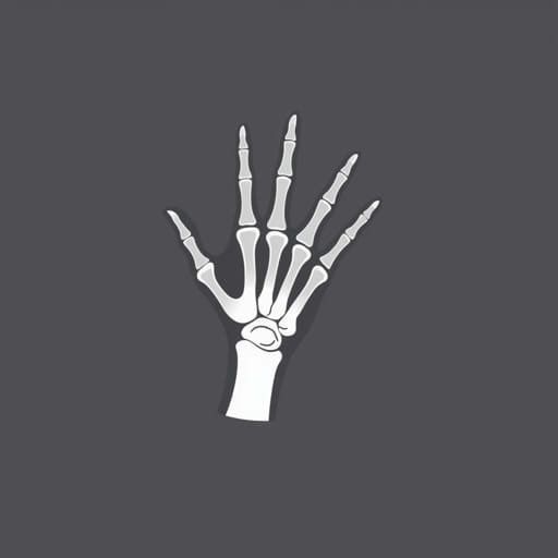
The hand oblique X-ray is an important diagnostic imaging technique used in medical practice to examine the bones and joints of the hand. This particular view offers an angled perspective, which helps in identifying subtle fractures, dislocations, or arthritic changes that may not be visible in standard views. The oblique position allows for better visualization of the metacarpals, phalanges, and intercarpal joints. It is commonly requested when patients present with pain, trauma, or suspected abnormalities that require detailed investigation. Understanding the purpose, technique, and interpretation of a hand oblique X-ray is crucial for healthcare professionals and patients alike.
What Is a Hand Oblique X-Ray?
A hand oblique X-ray is a type of radiographic image taken at an angle, usually around 45 degrees, from the standard posteroanterior (PA) or lateral views. This angled perspective provides a more detailed look at the hand’s bones and soft tissues. It is often performed as part of a complete hand series that includes the PA, lateral, and oblique views.
Purpose of the Oblique View
The primary reason for using an oblique view is to gain more information about the spatial relationships and overlapping structures of the hand. This view is especially helpful in identifying:
- Subtle fractures of the metacarpals and phalanges
- Joint space narrowing due to arthritis
- Bone lesions or infections
- Soft tissue swelling or abnormalities
- Alignment of bones following trauma
When Is a Hand Oblique X-Ray Recommended?
Physicians may recommend a hand oblique X-ray when a patient complains of:
- Pain in the fingers, knuckles, or wrist
- Swelling after trauma or injury
- Limited range of motion in the hand
- Signs of infection, such as redness or warmth
- Suspected foreign body embedded in the tissue
It is also used to evaluate healing after fractures or post-operative changes in the hand’s bone structure.
How the Procedure Is Performed
Getting a hand oblique X-ray is a simple and quick procedure that typically takes only a few minutes. Here’s how it usually works:
Patient Positioning
The patient is asked to sit beside or stand near the X-ray machine. The affected hand is placed on the X-ray table or platform at an oblique angle, typically with the palm resting at a 45-degree angle to the detector. The fingers are spread slightly to prevent overlapping.
Technician’s Role
The radiologic technologist ensures correct positioning and alignment. They might use a foam wedge or positioning tool to maintain the correct angle. The technician then steps away to take the X-ray while ensuring minimal exposure to radiation.
Safety Considerations
Though X-rays involve exposure to radiation, the amount used is minimal and considered safe for diagnostic purposes. Lead aprons may be used to shield other parts of the body if needed, especially in children or pregnant women.
Understanding the Results
Once the hand oblique X-ray is taken, it is reviewed by a radiologist a medical doctor trained to interpret imaging studies. The report may include:
- Presence or absence of fractures
- Evidence of degenerative changes like osteoarthritis
- Signs of infection such as bone erosion
- Bone alignment or displacement
- Condition of any surgical implants or healing fractures
Your primary care physician or specialist will then discuss the results and recommend treatment if necessary.
Advantages of the Oblique View
The oblique view offers several unique advantages over standard frontal and lateral views:
- Improved visualization of overlapping bones
- Enhanced detection of certain types of fractures
- Better assessment of the joint spaces
- More detailed anatomical information
These advantages make it a valuable tool in emergency departments, orthopedic clinics, and routine outpatient settings.
Common Conditions Diagnosed
Some of the most common conditions diagnosed using a hand oblique X-ray include:
- Boxer’s fracture (fracture of the fifth metacarpal)
- Osteoarthritis and rheumatoid arthritis
- Scaphoid fracture when extended to the hand
- Bone tumors or cysts
- Dislocations of the interphalangeal joints
In many cases, the oblique view reveals hidden injuries or changes not seen in the standard PA view alone.
Preparation and Aftercare
No special preparation is needed before a hand oblique X-ray. Patients should remove any jewelry or metal items from the hand and wrist. After the procedure, there is no recovery time needed, and patients can resume normal activities unless otherwise advised by their physician.
Follow-Up Imaging
If the X-ray shows unclear results or further detail is needed, the doctor may order additional imaging such as:
- CT scan for more detailed bone structure
- MRI for soft tissue injuries
- Ultrasound for tendon and ligament issues
These follow-up tests help build a complete picture of the hand’s condition.
Limitations of the X-Ray
While hand oblique X-rays are highly useful, they do have some limitations:
- Cannot visualize soft tissues as clearly as MRI
- May not detect hairline fractures in very early stages
- Not useful for functional or movement-related issues
Nevertheless, they are often the first step in diagnosis due to their accessibility and speed.
Hand oblique X-rays play an essential role in modern diagnostic radiology. By offering a detailed, angled view of the hand, they allow healthcare providers to detect injuries and abnormalities that might otherwise go unnoticed. Whether it’s evaluating trauma, arthritis, or a post-operative hand, this imaging technique provides vital information that guides patient care. If your doctor recommends a hand oblique X-ray, it is a safe and effective step toward understanding and treating your condition more accurately.
