The joint between the tarsus and the tibia and fibula is one of the most important weight-bearing joints in the human body. This joint plays a critical role in everyday movement such as walking, running, jumping, and standing. It connects the bones of the lower leg to the foot and enables essential actions like dorsiflexion and plantar flexion. Understanding the anatomy, structure, and function of this joint helps us appreciate how the foot and leg work together to support motion and balance.
Anatomy of the Ankle Joint
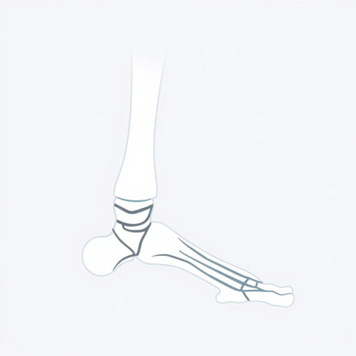
The joint formed between the tibia, fibula, and tarsus is commonly referred to as theankle jointor thetalocrural joint. It is a synovial hinge joint that allows movement primarily in one plane up and down which is crucial for locomotion and stability.
Main Bones Involved
- Tibia: The larger and medial bone of the lower leg. It bears the majority of the body’s weight and forms the top and medial side of the ankle joint.
- Fibula: The thinner, lateral bone of the lower leg. It provides lateral support to the ankle joint through the lateral malleolus.
- Talus: A tarsal bone located at the top of the foot. It sits under the tibia and fibula and forms the lower component of the ankle joint.
Joint Surfaces
The distal end of the tibia and the fibula form a socket called themortise, into which the superior surface of the talus fits snugly. This anatomical arrangement provides both stability and flexibility to the joint.
Ligaments Supporting the Joint
The joint between the tarsus and tibia-fibula is stabilized by several strong ligaments. These ligaments ensure that the bones stay aligned while allowing controlled motion.
Medial Ligament (Deltoid Ligament)
This is a strong, triangular-shaped ligament that supports the inside (medial side) of the ankle. It connects the tibia to the talus, calcaneus, and navicular bones of the tarsus.
Lateral Ligaments
- Anterior talofibular ligament (ATFL): Connects the fibula to the talus in front.
- Posterior talofibular ligament (PTFL): Connects the fibula to the talus behind.
- Calcaneofibular ligament: Connects the fibula to the calcaneus (heel bone).
Distal Tibiofibular Ligament Complex
This complex connects the tibia and fibula at the distal end, just above the ankle joint. It includes the anterior and posterior tibiofibular ligaments and the interosseous membrane. It helps maintain the mortise and supports the joint during weight-bearing.
Movements of the Joint
The joint between the tibia, fibula, and tarsus primarily allows for up-and-down movement of the foot. These are known as:
Dorsiflexion
Movement where the foot moves upward toward the shin. This motion occurs when walking uphill or lifting the toes off the ground.
Plantar Flexion
Movement where the foot points downward, such as when pressing a gas pedal or standing on tiptoes. The range of motion in plantar flexion is generally greater than in dorsiflexion.
Stability in Movement
While the primary motions are dorsiflexion and plantar flexion, the ankle joint works closely with surrounding joints in the foot to allow complex movements such as inversion, eversion, and rotation. These additional motions are supported by the subtalar and midtarsal joints.
Muscles Involved in Ankle Joint Movement
Several muscles cross the ankle joint to produce movement and provide stability. These muscles originate in the leg and insert in the foot, creating efficient motion at the joint.
Muscles for Dorsiflexion
- Tibialis anterior: Main dorsiflexor of the foot.
- Extensor hallucis longus: Lifts the big toe and assists in dorsiflexion.
- Extensor digitorum longus: Helps lift the smaller toes and dorsiflex the foot.
Muscles for Plantar Flexion
- Gastrocnemius: The prominent calf muscle that helps in powerful plantar flexion.
- Soleus: Works with the gastrocnemius to flex the foot downward.
- Flexor digitorum longus and flexor hallucis longus: Flex the toes and assist in plantar flexion.
Common Injuries at the Tibia-Fibula and Tarsus Joint
The ankle joint is one of the most commonly injured joints in the human body due to its weight-bearing function and range of motion. Injuries typically involve the ligaments, bones, or supporting tendons.
Ankle Sprains
These occur when the ligaments around the joint are stretched or torn. Lateral ankle sprains are most common and often involve the ATFL. Symptoms include swelling, bruising, and difficulty bearing weight.
Fractures
Fractures can involve the distal tibia, fibula, or talus. A common fracture is thePott’s fracture, which affects the lateral malleolus (distal fibula) and sometimes the medial malleolus (distal tibia). Fractures may result from direct trauma or twisting injuries.
Syndesmotic (High Ankle) Sprains
This injury affects the ligaments between the tibia and fibula, typically higher above the ankle. It is more severe than a typical ankle sprain and often occurs in athletes.
Arthritis
Chronic wear and tear or trauma can lead to arthritis in the ankle joint. Symptoms include pain, stiffness, swelling, and reduced mobility. Osteoarthritis is the most common form affecting this joint.
Diagnostic Imaging and Examination
In cases of injury or pain, clinical evaluation and imaging are essential to assess the joint. Common diagnostic tools include:
- X-rays: For viewing bone alignment and detecting fractures.
- MRI: For assessing soft tissue damage, ligament tears, or tendon injuries.
- CT scans: Useful for complex fractures or surgical planning.
Treatment and Rehabilitation
Treatment for ankle joint injuries depends on severity and may include both non-surgical and surgical options. Recovery aims to restore strength, motion, and stability to the joint.
Conservative Treatment
- Rest, Ice, Compression, Elevation (RICE)
- Physical therapy for range of motion and strength
- Bracing or casting for support and immobilization
- Anti-inflammatory medications for pain management
Surgical Treatment
Severe fractures, ligament ruptures, or chronic instability may require surgery. Procedures can include ligament repair, fracture fixation, or in advanced arthritis, ankle joint replacement or fusion.
The joint between the tarsus and the tibia-fibula, known as the ankle or talocrural joint, is a highly specialized and critical structure for mobility and weight-bearing. Its unique architecture, supported by bones, ligaments, and muscles, allows for efficient movement and stability of the lower limb. Awareness of its anatomy and common injuries is essential for athletes, clinicians, and anyone seeking to maintain healthy, functional movement. Proper care, strengthening, and timely treatment can ensure long-term joint health and injury prevention.