The right hepatic artery is one of the key vessels responsible for supplying oxygenated blood to the liver, specifically to its right lobe. Understanding the origin of the right hepatic artery is critical not only for anatomy students but also for surgeons and radiologists, especially during procedures such as liver resections, transplants, or laparoscopic cholecystectomy. Although the hepatic arterial system follows a general anatomical pattern, variations are common and can have clinical significance. The origin of the right hepatic artery, therefore, deserves close attention.
Standard Origin of the Right Hepatic Artery
Typical Anatomical Pathway
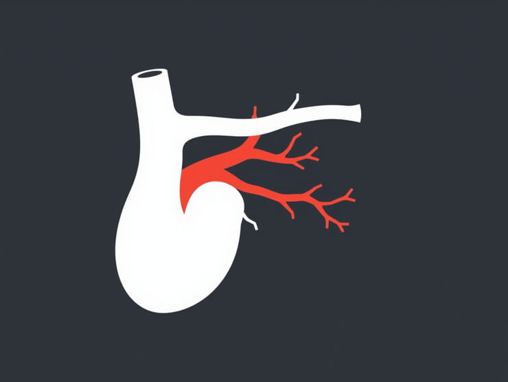
In the majority of individuals, the right hepatic artery originates as a branch of the proper hepatic artery. The proper hepatic artery itself is a continuation of the common hepatic artery, which arises from the celiac trunk a major branch of the abdominal aorta. After giving off the gastroduodenal artery, the common hepatic artery becomes the proper hepatic artery, which then typically bifurcates into the right and left hepatic arteries.
Location in the Hepatoduodenal Ligament
Once the right hepatic artery branches from the proper hepatic artery, it travels within the hepatoduodenal ligament a part of the lesser omentum along with the portal vein and common bile duct. It usually runs posterior to the common hepatic duct and enters the liver at the porta hepatis.
Branches and Supply
Functional Importance
The right hepatic artery supplies the right lobe of the liver, including the caudate lobe in many cases. It also gives rise to the cystic artery, which supplies the gallbladder. This makes the right hepatic artery especially important during gallbladder surgeries.
Key Branches
- Cystic artery supplies the gallbladder
- Segmental branches enter the liver and supply hepatic segments V to VIII
Anatomical Variations of the Right Hepatic Artery
Frequency of Variants
While the standard origin from the proper hepatic artery is common, anatomical studies show that up to 25% of individuals have variations in the origin of the right hepatic artery. These variations are important to identify preoperatively through imaging techniques like CT angiography or MR angiography.
Common Variants
- Replaced right hepatic artery from the superior mesenteric artery (SMA): This is the most common variation, where the right hepatic artery arises directly from the SMA instead of the proper hepatic artery.
- Accessory right hepatic artery from the SMA: In some cases, an accessory artery supplements the standard right hepatic artery rather than replacing it.
- Origin from the gastroduodenal artery or directly from the common hepatic artery: Less common but documented in anatomical studies.
Terminology
It is important to distinguish between a replaced right hepatic artery and an accessory artery:
- Replaced artery: the sole arterial supply to the region, replacing the usual source.
- Accessory artery: an additional source of blood supply alongside the standard branch.
Embryological Basis of Variation
Development of Hepatic Arteries
The variations in the origin of the right hepatic artery can be explained by the complex embryological development of the abdominal arteries. During fetal development, multiple anastomoses form between the vitelline arteries, which eventually give rise to branches such as the celiac trunk and SMA. Persistence or regression of different embryonic channels can result in variations in the adult arterial anatomy.
Clinical Significance
Surgical Implications
Knowing the origin of the right hepatic artery is crucial in liver and biliary surgeries. Unrecognized variants can lead to accidental injury, resulting in liver ischemia or bile duct damage. This is especially true during:
- Laparoscopic cholecystectomy
- Liver transplantation
- Hepatectomy or segmental liver resections
Imaging and Preoperative Planning
Radiologists play an important role in identifying hepatic artery variations using modern imaging modalities. Preoperative angiography helps map the vascular anatomy to minimize complications during surgical intervention.
Interventional Procedures
The right hepatic artery may also be targeted in interventional radiology procedures, such as:
- Chemoembolization for liver tumors (TACE)
- Embolization of bleeding hepatic vessels
- Stent placement or angioplasty
Right Hepatic Artery and Liver Segmentation
Couinaud’s Liver Segments
According to Couinaud’s classification, the liver is divided into eight functional segments. The right hepatic artery predominantly supplies:
- Segment V (anterior inferior)
- Segment VI (posterior inferior)
- Segment VII (posterior superior)
- Segment VIII (anterior superior)
Segmental Branching
After entering the liver parenchyma, the right hepatic artery branches further to supply these segments. Segmental arterial supply is often used to guide resection during liver surgery to preserve as much functional tissue as possible.
Comparative Anatomy and Variants in Populations
Ethnic and Geographic Differences
Some studies suggest that the prevalence of replaced or accessory right hepatic arteries may differ across ethnic groups or populations. For example:
- Higher incidence of replaced right hepatic artery in Asian populations
- More frequent accessory arteries reported in cadaveric studies in European populations
These differences highlight the importance of individual assessment rather than relying solely on textbook anatomy.
Summary of Key Points
- The right hepatic artery usually originates from the proper hepatic artery, a branch of the common hepatic artery from the celiac trunk.
- It courses through the hepatoduodenal ligament and supplies the right lobe of the liver and the gallbladder.
- Variants include origin from the superior mesenteric artery, which may be replaced or accessory.
- Embryological development explains many of the observed anatomical variations.
- Identification of the artery’s origin is vital for safe surgical and interventional procedures.
Understanding the origin of the right hepatic artery is fundamental in both anatomical education and clinical practice. While most individuals have a right hepatic artery arising from the proper hepatic artery, variations are not rare and carry significant implications. Surgeons, radiologists, and medical students must be familiar with both the standard and variant anatomy to avoid complications during procedures involving the liver and biliary tract. Accurate knowledge of the right hepatic artery’s origin, course, and branches ensures better surgical outcomes and patient safety, making it an essential focus in hepatobiliary anatomy.