The common peroneal nerve, also known as the common fibular nerve, is one of the key nerves in the lower limb that plays an essential role in leg movement and sensation. It branches from the sciatic nerve and provides motor and sensory innervation to specific parts of the leg and foot. Understanding the common peroneal nerve innervation is vital for healthcare professionals, anatomy students, and anyone interested in how the nervous system supports movement, balance, and coordination. Damage or compression of this nerve can lead to serious complications such as foot drop or sensory loss, making its anatomy and function particularly important to study.
Anatomy and Course of the Common Peroneal Nerve
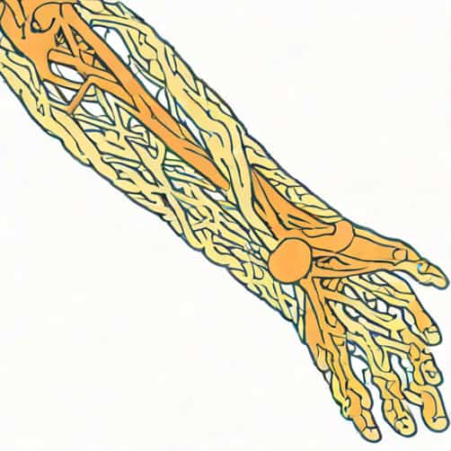
The common peroneal nerve originates as one of the two terminal branches of the sciatic nerve, the other being the tibial nerve. It typically arises in the upper part of the popliteal fossa, which is the shallow depression located behind the knee joint. From there, the nerve travels laterally along the border of the biceps femoris muscle before wrapping around the neck of the fibula, where it becomes superficial and vulnerable to injury.
After winding around the fibular neck, the common peroneal nerve divides into two major terminal branches
- The superficial peroneal (fibular) nerveresponsible primarily for motor supply to the muscles of the lateral compartment of the leg and sensory innervation to most of the dorsum of the foot.
- The deep peroneal (fibular) nerveresponsible for motor supply to the muscles of the anterior compartment of the leg and sensory innervation to the web space between the first and second toes.
Throughout its course, the common peroneal nerve also gives off smaller branches, including articular branches to the knee joint and a cutaneous branch known as the lateral sural cutaneous nerve, which provides sensation to the upper lateral surface of the leg.
Motor Innervation of the Common Peroneal Nerve
The motor function of the common peroneal nerve is carried out through its two major branches. These branches control several key muscles involved in foot movement and ankle stability.
1. Superficial Peroneal Nerve Motor Function
The superficial peroneal nerve supplies the two muscles in the lateral compartment of the leg
- Fibularis longus (peroneus longus)Responsible for eversion of the foot and assistance in plantarflexion of the ankle. It also helps maintain the arch of the foot.
- Fibularis brevis (peroneus brevis)Also contributes to foot eversion and stabilization during walking.
These muscles work together to turn the sole of the foot outward and prevent the foot from rolling inward excessively, an action that is important for balance and movement on uneven surfaces.
2. Deep Peroneal Nerve Motor Function
The deep peroneal nerve innervates the muscles of the anterior compartment of the leg, which are primarily responsible for dorsiflexion of the ankle and extension of the toes. These include
- Tibialis anteriorDorsiflexes and inverts the foot, helping to lift the foot during walking.
- Extensor hallucis longusExtends the big toe and assists in dorsiflexion of the foot.
- Extensor digitorum longusExtends the other toes and assists in dorsiflexion.
- Fibularis tertiusA small muscle that helps in dorsiflexion and eversion of the foot.
These muscles are crucial for normal gait, as they allow the foot to clear the ground during the swing phase of walking and control the lowering of the foot to the ground after heel strike.
Sensory Innervation of the Common Peroneal Nerve
In addition to motor control, the common peroneal nerve provides sensory innervation to parts of the leg and foot. Sensory branches of this nerve are responsible for carrying information about touch, pain, and temperature from specific areas of the skin to the brain.
- Lateral sural cutaneous nerveArises directly from the common peroneal nerve and supplies sensation to the upper lateral surface of the leg.
- Superficial peroneal nerveProvides sensory innervation to most of the dorsum of the foot, except the web space between the first and second toes.
- Deep peroneal nerveSupplies sensory innervation to the skin between the first and second toes, an area often referred to as the flip-flop space.
This distribution ensures that the lateral and anterior aspects of the leg, as well as the top of the foot, receive sensory input, which is important for maintaining balance and spatial awareness during movement.
Common Peroneal Nerve Injury
The common peroneal nerve is one of the most commonly injured nerves in the lower limb because of its superficial position around the neck of the fibula. Injury can result from trauma, prolonged pressure, or medical conditions affecting nerve function.
Causes of Injury
- Fracture or dislocation of the fibular head or neck
- Prolonged leg crossing or tight casts and braces
- Direct trauma during sports or accidents
- Compression due to weight loss or prolonged immobility
- Diabetes-related neuropathy or systemic nerve disorders
Symptoms of Common Peroneal Nerve Damage
When the common peroneal nerve is injured, it can cause both motor and sensory deficits. The most characteristic symptom is foot drop, where the patient is unable to lift the front part of the foot due to paralysis of the dorsiflexor muscles. This results in a steppage gait, where the person lifts their knee higher than usual to avoid dragging the toes.
Other symptoms may include
- Numbness or tingling along the lateral side of the leg and top of the foot
- Weakness in foot eversion
- Loss of sensation between the first and second toes
- Pain or burning sensation over the fibular neck
Diagnosis and Testing
Diagnosis of common peroneal nerve injury involves a combination of physical examination and diagnostic testing. A doctor may assess muscle strength, reflexes, and sensory responses in the affected limb. Nerve conduction studies and electromyography (EMG) can help measure the electrical activity of the muscles and identify the location and severity of the nerve damage.
Imaging techniques like MRI or ultrasound may be used to identify structural causes such as compression, swelling, or bone injury affecting the nerve pathway.
Treatment and Recovery
Treatment for common peroneal nerve damage depends on the underlying cause and severity of the injury. In many cases, non-surgical management can help restore function and prevent further damage.
Non-Surgical Treatments
- Physical therapyStrengthening and stretching exercises help maintain muscle tone and improve mobility.
- BracingAn ankle-foot orthosis (AFO) can be used to support the foot and correct foot drop.
- MedicationPain relievers or anti-inflammatory drugs may help reduce discomfort and inflammation.
- Activity modificationAvoiding prolonged pressure on the fibular head, such as crossing the legs or tight clothing, can prevent worsening of symptoms.
Surgical Treatments
In cases where the nerve is severely compressed or damaged, surgical intervention may be necessary. Options include decompression surgery to relieve pressure on the nerve, or nerve grafting and repair for more extensive injuries. Postoperative rehabilitation is essential to regain strength and movement.
Prevention and Care
Since the common peroneal nerve is superficial near the fibular neck, prevention focuses on avoiding external compression and maintaining good leg posture. Using padding during long surgeries, avoiding tight casts, and maintaining proper sitting posture can help protect this nerve. For athletes, strengthening the muscles around the knee and ankle reduces the risk of nerve traction injuries during sports activities.
The common peroneal nerve innervation plays a crucial role in both movement and sensation of the lower leg and foot. Its branches the superficial and deep peroneal nerves coordinate essential actions such as dorsiflexion, eversion, and toe extension, while providing sensory feedback from the skin. Because of its superficial location, the nerve is prone to injury, leading to conditions like foot drop or sensory loss. Understanding its anatomy, functions, and clinical relevance helps in preventing injury and ensuring proper diagnosis and treatment when problems arise. The common peroneal nerve truly represents a key connection between the nervous system and the mechanics of lower limb movement.