The maxillary first molar is one of the most significant teeth in the human dentition due to its role in chewing, occlusion, and overall oral health. Understanding its anatomy, particularly the number of roots it typically possesses, is essential for dental professionals in performing procedures such as root canal therapy, extractions, and restorative dentistry. The number and configuration of roots in maxillary first molars can vary, making it an important topic in endodontics and dental anatomy education. Proper knowledge of this tooth’s root anatomy is crucial to prevent treatment complications and ensure successful dental interventions.
Anatomy of the Maxillary First Molar
The maxillary first molar is located in the upper jaw, behind the second premolar, and is usually the first permanent molar to erupt, typically around the age of six. It is characterized by a broad occlusal surface with multiple cusps and a complex root structure. The morphology of this tooth makes it highly functional for grinding and mastication, contributing to overall dental occlusion.
Cusps and Occlusal Features
The maxillary first molar generally has four major cusps the mesiobuccal, distobuccal, mesiolingual, and distolingual cusps. Some molars may also have a smaller fifth cusp known as the cusp of Carabelli on the mesiolingual side. The occlusal anatomy, combined with the root structure, affects the tooth’s stability and anchorage in the jaw.
Number of Roots in Maxillary First Molar
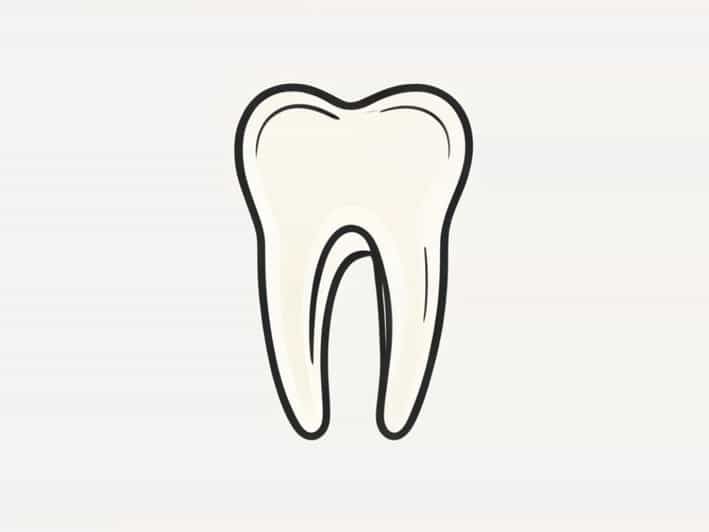
Typically, the maxillary first molar has three roots. These roots are usually distinct and positioned to provide maximum stability and support for the tooth
- Mesiobuccal RootLocated on the front outer side of the tooth, it often contains two canals, though sometimes only one is present. The complexity of the mesiobuccal root makes it a focus in endodontic procedures.
- Distobuccal RootPositioned on the back outer side, usually containing a single canal. This root is narrower than the mesiobuccal root and is typically easier to treat during root canal therapy.
- Palatal RootThe largest and strongest root, located on the inner side of the tooth toward the palate. It generally contains a single, large canal and provides significant anchorage for the tooth.
Variations in Root Number
While three roots are most common, anatomical variations can occur. Occasionally, a maxillary first molar may have four roots, especially if an extra root develops on the mesiobuccal or distobuccal side. Conversely, some molars may have fused roots, resulting in only two visible roots. Understanding these variations is critical for dental procedures, as missing a canal or misidentifying root morphology can lead to treatment failure.
Root Canal Anatomy and Clinical Implications
The internal canal structure of the maxillary first molar is as important as the number of roots. Each root typically contains one or more canals
- Mesiobuccal RootUsually has two canals, termed MB1 and MB2. The MB2 canal can be difficult to locate, making thorough examination essential during root canal therapy.
- Distobuccal RootUsually has a single canal, which is relatively straight and easier to treat.
- Palatal RootTypically has one large, curving canal that may present challenges if not carefully navigated.
Failure to recognize additional canals, particularly in the mesiobuccal root, can compromise endodontic outcomes. Modern imaging techniques, such as cone-beam computed tomography (CBCT), are increasingly used to identify root canal morphology accurately before treatment.
Clinical Significance of Root Number
Knowing the typical number of roots and their variations is critical in several dental practices
Endodontic Treatment
Root canal therapy requires precise cleaning, shaping, and filling of all canals. The presence of multiple roots and canals in maxillary first molars makes the procedure more complex. Identifying all roots and canals reduces the risk of untreated canals and subsequent infection.
Extractions and Surgical Procedures
During extractions or apicoectomy procedures, awareness of root number and position prevents accidental damage to surrounding bone and adjacent teeth. The palatal root, being the largest, may require careful manipulation to avoid fracture.
Restorative Dentistry
When placing crowns, bridges, or implants, understanding the root anatomy ensures adequate support and prevents complications. Proper evaluation of root number and structure helps dentists plan restorative procedures that maintain long-term tooth stability.
Factors Affecting Root Morphology
Several factors influence the number and configuration of roots in maxillary first molars
- Genetic FactorsIndividual genetic variation can lead to differences in root number and shape.
- Developmental AnomaliesSometimes, roots may fuse or split abnormally during tooth development.
- Ethnic DifferencesStudies show variations in root morphology across populations, affecting treatment approaches.
- Age and WearOver time, roots may undergo resorption or changes due to periodontal disease, affecting their clinical appearance.
Diagnostic Tools for Root Assessment
Accurate assessment of root number and morphology is essential before dental procedures. Common diagnostic tools include
- Periapical RadiographsTraditional X-rays provide two-dimensional images of roots but may not reveal complex canal systems.
- Panoramic RadiographsOffer a broader view of the maxilla, useful for planning extractions or implants.
- Cone-Beam Computed Tomography (CBCT)Provides detailed three-dimensional images of root structures, allowing precise identification of all roots and canals.
- Endodontic MicroscopesMagnification aids in locating additional canals, especially in the mesiobuccal root.
The maxillary first molar typically has three roots the mesiobuccal, distobuccal, and palatal roots, each with its unique anatomy and clinical considerations. Variations, such as extra roots or fused roots, can occur and have significant implications for endodontic treatment, extractions, and restorative procedures. Understanding the root number, canal configuration, and possible variations is crucial for dental professionals to ensure successful outcomes. With modern diagnostic tools and careful examination, clinicians can navigate the complex anatomy of the maxillary first molar and provide effective, precise, and safe dental care. Proper knowledge of this tooth’s root system remains a fundamental aspect of dental education and clinical practice, emphasizing the importance of anatomy in everyday dentistry.