Electrocardiography, often abbreviated as ECG or EKG, is a medical procedure that records the electrical activity of the heart over a period of time. It is a non-invasive and widely used diagnostic tool that provides essential information about the heart’s rhythm, structure, and overall health. By analyzing the electrical signals generated by the heart muscles during contraction and relaxation, healthcare providers can detect abnormalities, diagnose heart conditions, and monitor the effects of treatments or medications. Electrocardiography plays a crucial role in cardiology, emergency medicine, and routine health checkups, making it one of the most important tools for assessing heart function in both clinical and outpatient settings.
Understanding Electrocardiography
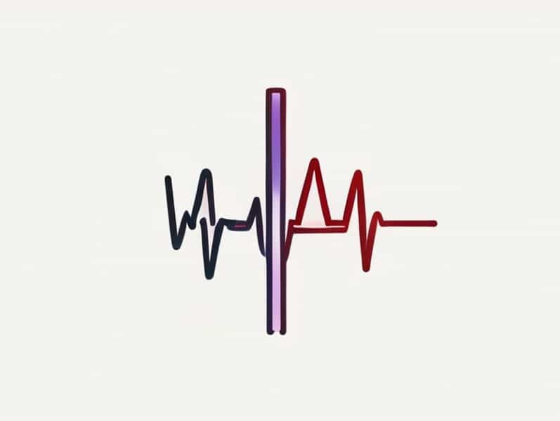
Electrocardiography involves the placement of electrodes on a patient’s skin to capture the heart’s electrical signals. These signals are then recorded and displayed as a waveform, known as an electrocardiogram. The electrocardiogram provides visual representation of the heart’s electrical activity, showing the sequence of depolarization and repolarization of the atria and ventricles. By examining the various components of the ECG waveform, such as the P wave, QRS complex, and T wave, medical professionals can assess whether the heart is functioning normally or if there are irregularities that require further investigation.
Purpose of Electrocardiography
The primary purpose of electrocardiography is to detect and diagnose conditions affecting the heart. It is commonly used to
- Identify arrhythmias, which are irregular heartbeats that can lead to serious complications if untreated.
- Detect myocardial infarction (heart attack) by revealing characteristic changes in the ECG pattern.
- Assess structural abnormalities, such as enlarged chambers or hypertrophy of the heart walls.
- Monitor the effectiveness of treatments for heart diseases, including medications or pacemaker therapy.
- Evaluate the effects of electrolyte imbalances or other metabolic conditions on heart function.
How Electrocardiography Works
Electrocardiography works by detecting tiny electrical impulses generated by the heart as it beats. These impulses travel through the heart muscles and are conducted to the body’s surface, where they can be measured using electrodes. Typically, multiple electrodes are placed on the chest, arms, and legs to obtain a comprehensive view of the heart’s electrical activity from different angles. The recorded signals are then displayed on a monitor or printed on paper as an electrocardiogram, which can be analyzed for patterns that indicate normal or abnormal cardiac activity.
Components of an ECG
An electrocardiogram consists of several key components, each representing a specific phase of the heart’s electrical cycle
- P waveRepresents atrial depolarization, which is the electrical activity that triggers atrial contraction.
- QRS complexCorresponds to ventricular depolarization, the electrical event that initiates ventricular contraction.
- T waveIndicates ventricular repolarization, which is the process of the ventricles returning to their resting electrical state.
- PR intervalMeasures the time between the onset of atrial depolarization and the start of ventricular depolarization.
- ST segmentRepresents the period when the ventricles are depolarized and preparing to repolarize, important for detecting myocardial ischemia or injury.
Types of Electrocardiography
Electrocardiography can be performed in several forms, depending on the clinical requirements and the duration of monitoring needed. The common types include
Resting ECG
Resting ECG is performed while the patient is lying down in a relaxed state. It is the standard procedure for routine heart evaluation and provides a quick assessment of heart rhythm and electrical activity.
Exercise or Stress ECG
Stress ECG, also called a treadmill test, records the heart’s activity during physical exertion. This test helps identify issues such as coronary artery disease that may not be apparent when the heart is at rest. It is particularly useful in evaluating chest pain, shortness of breath, or other exertion-related symptoms.
Holter Monitoring
Holter monitoring involves wearing a portable ECG device for 24 to 48 hours or longer. This continuous monitoring captures variations in heart rhythm that may occur intermittently and could be missed by a standard resting ECG. It is especially helpful for detecting sporadic arrhythmias or unexplained palpitations.
Event Monitoring
Event monitors are used for long-term ECG recording, typically over weeks or months. Patients activate the device when they experience symptoms, allowing healthcare providers to correlate the electrical activity with specific events or sensations. This method is effective in diagnosing infrequent cardiac episodes that are difficult to capture otherwise.
Applications of Electrocardiography
Electrocardiography has a wide range of applications in medical practice, making it an indispensable diagnostic tool for cardiologists and other healthcare professionals. Some common applications include
- Routine health checkups to assess overall heart function.
- Diagnosis and monitoring of heart rhythm disorders, including atrial fibrillation and tachycardia.
- Detection of heart attacks and ischemic conditions through characteristic ECG changes.
- Evaluation of congenital heart defects and structural abnormalities.
- Monitoring the impact of medications that affect cardiac function.
- Pre-operative assessment before surgery to ensure heart safety.
Benefits of Electrocardiography
Electrocardiography offers numerous benefits due to its non-invasive nature, ease of use, and informative results. Key advantages include
- Provides immediate insights into the heart’s electrical activity.
- Helps in early detection of life-threatening conditions such as heart attacks.
- Facilitates monitoring and management of chronic heart diseases.
- Is safe, painless, and requires minimal preparation.
- Assists healthcare providers in making informed decisions about treatment and interventions.
Limitations of Electrocardiography
While ECG is highly valuable, it has certain limitations that healthcare providers consider when interpreting results. An ECG provides a snapshot of the heart’s electrical activity at a given moment, which means it may not detect intermittent problems unless continuous monitoring is employed. It also may not reveal structural heart issues such as valve defects or minor blockages in coronary arteries, which often require additional imaging tests like echocardiography or angiography. Despite these limitations, ECG remains a fundamental tool in cardiovascular diagnosis and monitoring.
Electrocardiography is a fundamental medical procedure that provides essential information about the heart’s electrical activity. It helps in diagnosing arrhythmias, heart attacks, structural abnormalities, and other cardiovascular conditions. Through resting ECG, stress tests, Holter monitoring, and event monitoring, healthcare providers can obtain comprehensive insights into a patient’s cardiac health. Its non-invasive nature, ease of use, and informative results make electrocardiography one of the most important diagnostic tools in modern medicine. Understanding what electrocardiography means and how it is performed allows patients to appreciate the value of this procedure in maintaining heart health and preventing potential complications.