Electrophoresis is a widely used laboratory technique that separates charged molecules, such as DNA, RNA, or proteins, based on their size and electrical charge. This method relies on the principle that charged ptopics move through a medium when an electric field is applied. Understanding electrophoresis and its diagrammatic representation is essential for students, researchers, and professionals in molecular biology, genetics, and biochemistry. The technique not only helps visualize biomolecules but also enables their analysis, purification, and quantification for a variety of scientific applications.
Principle of electrophoresis
The fundamental principle of electrophoresis is that charged molecules migrate toward an electrode with opposite charge under the influence of an electric field. Negatively charged molecules, such as DNA and RNA, move toward the positive electrode (anode), while positively charged molecules move toward the negative electrode (cathode). The rate of migration depends on factors such as the molecule’s size, shape, and charge, as well as the composition and density of the medium through which it moves.
Diagrammatic representation of electrophoresis
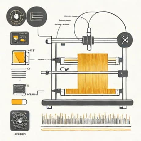
A simple diagrammatic representation of electrophoresis typically includes the following components
- Electrophoresis gelUsually made of agarose or polyacrylamide, providing a matrix for separation.
- Sample wellsSlots at one end of the gel where DNA, RNA, or protein samples are loaded.
- Buffer solutionConducts electricity and maintains pH during the run.
- ElectrodesNegative (cathode) and positive (anode) electrodes connected to a power supply.
- Migration pathMolecules move from the wells toward the electrode with opposite charge.
In a diagram, DNA fragments typically appear as bands at different positions along the gel after staining. Smaller fragments move faster and appear closer to the positive electrode, while larger fragments migrate more slowly. A marker or ladder is often run alongside samples to provide size references.
Types of electrophoresis
Electrophoresis can be performed using different media and methods, depending on the molecules being analyzed. The two most common types are agarose gel electrophoresis and polyacrylamide gel electrophoresis (PAGE).
Agarose gel electrophoresis
Agarose gels are primarily used for separating DNA and RNA fragments. The concentration of agarose in the gel determines the pore size, which affects the resolution of molecules of different lengths. Agarose gel electrophoresis is widely used in genetic research, DNA cloning, and PCR product analysis. DNA fragments are visualized using dyes such as ethidium bromide or safer alternatives under UV light.
Polyacrylamide gel electrophoresis (PAGE)
PAG E provides higher resolution and is used for separating smaller DNA fragments, RNA, and proteins. Proteins are often analyzed using SDS-PAGE, where sodium dodecyl sulfate (SDS) denatures proteins and provides a uniform negative charge. This allows separation based solely on size. PAGE can be further enhanced using gradient gels, which improve resolution of complex protein mixtures.
Applications of electrophoresis
Electrophoresis has numerous applications in molecular biology, genetics, biochemistry, and medical research. Its versatility makes it an essential tool for analyzing nucleic acids, proteins, and other biomolecules. Some of the key applications include
DNA and RNA analysis
Electrophoresis is widely used to separate and analyze DNA and RNA fragments. Applications include
- Determining the size of PCR products or restriction enzyme digests
- Checking the integrity of RNA samples for downstream experiments
- Genotyping organisms and identifying genetic variations
- Performing DNA fingerprinting for forensic and paternity testing
Protein analysis
Protein electrophoresis enables researchers to study the composition, size, and quantity of proteins. Common applications include
- SDS-PAGE for size-based protein separation
- Western blotting, where proteins are transferred to a membrane and probed with specific antibodies
- Isoelectric focusing to separate proteins based on their isoelectric point
- Analyzing enzyme activity and post-translational modifications
Medical diagnostics
Electrophoresis plays a critical role in medical diagnostics. Examples include
- Hemoglobin electrophoresis to diagnose sickle cell anemia and thalassemia
- Serum protein electrophoresis to detect multiple myeloma or other plasma disorders
- Viral RNA analysis for pathogen detection and monitoring infection
Research and biotechnology
In research and biotechnology, electrophoresis is indispensable for
- Cloning and sequencing DNA fragments
- Purifying nucleic acids and proteins for functional studies
- Studying protein-protein interactions and molecular complexes
- Monitoring gene expression and transcriptional activity
Advantages of electrophoresis
Electrophoresis offers several advantages that make it a preferred method for molecular analysis
- High resolution for separating molecules of different sizes
- Ability to analyze complex mixtures of DNA, RNA, or proteins
- Relatively simple and cost-effective technique
- Compatibility with downstream applications such as blotting or sequencing
- Provides visual representation of molecular size and abundance
Limitations and considerations
Despite its advantages, electrophoresis has some limitations. It requires careful handling to avoid sample contamination, and visualization methods may involve toxic chemicals like ethidium bromide. The resolution may be limited for very small or very large molecules, and interpretation of results requires experience. Additionally, the process can be time-consuming for complex samples. Advances in technology, however, continue to improve sensitivity, safety, and automation.
Electrophoresis is a fundamental technique in molecular biology and biochemistry, used to separate and analyze DNA, RNA, and proteins based on size and charge. Diagrammatically, it involves a gel matrix, sample wells, buffer, and electrodes, allowing molecules to migrate under an electric field and form visible bands. Its applications span DNA analysis, protein studies, medical diagnostics, and research, making it an invaluable tool for scientists. Despite some limitations, the method’s versatility, accuracy, and visual output make it one of the most widely employed techniques in laboratories worldwide, providing insights into molecular structure, function, and interactions.