The trigeminal nerve maxillary branch, also known as the second division of the trigeminal nerve or V2, is a crucial component of the human nervous system responsible for transmitting sensory information from the midfacial region to the brain. This branch plays a vital role in sensations such as touch, pain, and temperature from areas including the lower eyelid, upper lip, cheeks, nasal cavity, and upper teeth. Understanding the anatomy, function, clinical significance, and potential disorders related to the trigeminal nerve maxillary branch is essential for medical professionals, dental specialists, and students of neurology and anatomy, as it impacts both diagnosis and treatment strategies for facial pain and sensory disorders.
Anatomy of the Maxillary Branch
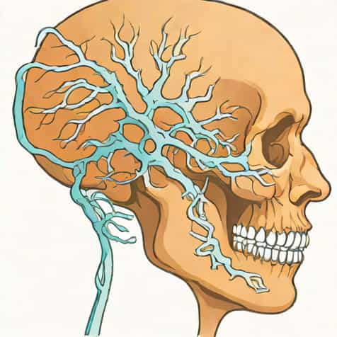
The maxillary branch of the trigeminal nerve emerges from the trigeminal ganglion, located in the Meckel’s cave of the cranial cavity. Unlike the mandibular branch, the maxillary division is purely sensory and does not carry motor fibers. It travels through the foramen rotundum, entering the pterygopalatine fossa, where it gives off several branches before reaching the face. The anatomical course of the maxillary branch is essential for clinicians performing nerve blocks or surgical interventions in the midface region to avoid complications.
Key Anatomical Features
- Originates from the trigeminal ganglion
- Passes through the foramen rotundum into the pterygopalatine fossa
- Purely sensory, no motor fibers
- Divides into several branches, including infraorbital, zygomatic, and superior alveolar nerves
- Innervates midfacial regions including upper lip, cheeks, nasal cavity, and upper teeth
Branches and Distribution
The maxillary branch gives rise to several important nerves that provide sensory innervation to the midface. The infraorbital nerve continues along the infraorbital groove and canal, supplying the lower eyelid, upper lip, and lateral nose. The zygomatic nerve branches further into the zygomaticotemporal and zygomaticofacial nerves, innervating the lateral forehead and cheek. Superior alveolar nerves, including anterior, middle, and posterior branches, innervate the upper teeth and gingiva. Understanding these branches is critical for dental procedures, regional anesthesia, and managing facial pain syndromes.
Major Branches and Functions
- Infraorbital nerve sensation to lower eyelid, upper lip, lateral nose
- Zygomatic nerve lateral forehead and cheek sensation
- Posterior superior alveolar nerve upper molar teeth and gingiva
- Middle superior alveolar nerve upper premolar teeth and gingiva
- Anterior superior alveolar nerve upper incisor and canine teeth, anterior maxillary gingiva
- Nasopalatine and greater palatine nerves sensation to the hard palate and anterior nasal cavity
Function of the Maxillary Branch
The primary function of the trigeminal nerve maxillary branch is to convey sensory information from the midfacial region to the central nervous system. It allows individuals to perceive touch, pressure, pain, and temperature. The branch also plays a role in reflexes such as blinking and facial protective responses by relaying information about potential threats or irritants. Its purely sensory function distinguishes it from the mandibular branch, which contains both motor and sensory fibers. Proper function is essential for normal facial sensation, oral health, and coordination of protective reflexes.
Sensory Functions
- Touch and pressure perception in midface
- Pain detection in upper teeth, palate, and midface
- Temperature sensation in cheek, upper lip, and nasal cavity
- Contribution to protective reflexes such as blinking
- Facilitation of oral and dental sensory feedback
Clinical Significance
The maxillary branch of the trigeminal nerve is clinically significant in both diagnostics and therapeutic procedures. Disorders affecting this nerve can result in numbness, tingling, or pain in its distribution area. Trigeminal neuralgia, particularly involving the maxillary branch, causes severe, paroxysmal facial pain often triggered by light touch, chewing, or speaking. Accurate identification of the nerve’s anatomy is also crucial for local anesthesia in dental procedures, maxillofacial surgery, and the treatment of sinus or nasal cavity conditions. Understanding variations and common anatomical landmarks ensures effective treatment while minimizing complications.
Clinical Applications
- Trigeminal nerve blocks for anesthesia in dental and surgical procedures
- Management of trigeminal neuralgia affecting V2 distribution
- Diagnosis of facial sensory deficits following trauma or surgery
- Evaluation of sinus infections or tumors impacting the maxillary branch
- Use in reconstructive and cosmetic maxillofacial procedures
Common Disorders and Complications
Several conditions may affect the trigeminal nerve maxillary branch. Trigeminal neuralgia is characterized by sudden, severe facial pain, often localized to the upper teeth, cheeks, or upper lip. Herpes zoster infection can lead to postherpetic neuralgia, causing persistent pain along the nerve distribution. Trauma, fractures of the maxilla or orbit, and surgical complications can result in numbness or sensory loss. Early diagnosis and management are essential to prevent chronic pain or permanent sensory deficits. Treatment may involve medications, nerve blocks, or surgical interventions depending on the severity and underlying cause.
Examples of Disorders
- Trigeminal neuralgia (V2 involvement)
- Postherpetic neuralgia following shingles
- Trauma-induced sensory loss or paresthesia
- Maxillofacial fractures affecting nerve function
- Complications from dental surgery or sinus procedures
Diagnostic and Therapeutic Approaches
Diagnosis of maxillary branch disorders typically involves a combination of clinical examination, patient history, and imaging techniques such as MRI or CT scans. Neurological assessments test sensory perception, reflexes, and pain response within the V2 distribution. Therapeutic approaches may include pharmacological management with anticonvulsants or analgesics, nerve blocks for acute pain relief, and, in severe cases, surgical decompression or rhizotomy. Dental practitioners use local anesthesia targeting the infraorbital nerve for procedures on the upper teeth, providing effective pain control while minimizing systemic effects.
Diagnostic Methods
- Neurological examination of midface sensory function
- Imaging MRI, CT scans for structural evaluation
- Electrophysiological testing for nerve conduction
- Pain mapping to identify specific V2 involvement
- Dental examination for upper teeth and gingiva sensory feedback
Treatment Strategies
- Anticonvulsants or analgesics for trigeminal neuralgia
- Nerve blocks or local anesthesia for dental or surgical procedures
- Surgical interventions in refractory or structural cases
- Physical therapy and pain management techniques
- Post-operative monitoring to prevent long-term sensory deficits
The trigeminal nerve maxillary branch is a vital sensory component of the nervous system, responsible for transmitting critical information from the midface to the brain. Its complex anatomy, distribution, and clinical significance make it essential for dental, neurological, and maxillofacial practice. Disorders affecting this nerve, including trigeminal neuralgia, postherpetic neuralgia, and trauma-related sensory loss, highlight the importance of accurate diagnosis and effective treatment strategies. A thorough understanding of the trigeminal nerve maxillary branch ensures proper management of facial pain, improved outcomes in surgical procedures, and preservation of normal facial sensation, contributing to both patient comfort and clinical success.